The Lenses Through Which the Brain Speaks: Understanding EEG Montages
 When Hans Berger first recorded the alpha rhythm, he did something deceptively simple: he placed electrodes on the scalp and compared the voltage between them.
Two points.
One difference.
A wave.
That is the essence of EEG — every line on the screen is nothing more than the electrical difference between two places on the scalp.
But how we pair those electrodes, how we arrange them, how we “listen” — that changes everything.
Imagine trying to understand a complex symphony using microphones placed at different corners of a concert hall.
Move the microphones, and the sound changes.
Place them closer together, and subtle harmonics emerge.
Spread them apart, and broad reverberations dominate.
The music is the same, but the perspective alters the meaning.
This is the world of EEG montages — the art of choosing the right arrangement of electrodes to reveal the truth hidden in the cortex.
What Is a Montage? A Viewpoint, Not a Trick
A montage is simply the pattern in which EEG channels are displayed — the specific pairings of electrodes used to generate each trace.
The brain is unchanged.
The patient is unchanged.
But the interpretation shifts drastically depending on which montage you use.
A montage is the brain seen through a particular lens.
Some lenses sharpen focal abnormalities.
Some highlight symmetry.
Some reveal hidden spikes that other lenses conceal.
Like a radiologist choosing CT slices, an EEG reader must know which montage reveals which truth.
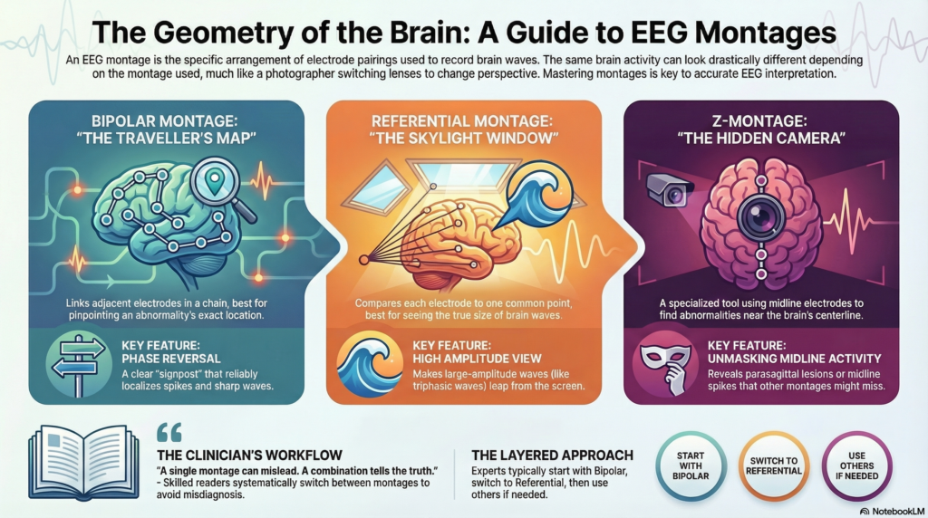
1. The Bipolar Montage — The Traveller’s Map
Shown clearly on pages 4–5 of the handbook [1], the bipolar montage links each electrode to its neighbour in a chain-like fashion.
Fp1 → F3
F3 → C3
C3 → P3
P3 → O1
Each pair forms a channel.
Each channel shows the difference between two adjacent electrodes.
The bipolar montage feels like walking across the scalp — step by step, lead by lead, region by region.
It is breathtakingly useful for one reason:
Phase Reversal — The EEG Compass
If a spike or sharp wave is real, the electrical field reverses direction at the point where the abnormality is strongest.
This creates a phase reversal, a tell-tale flip in polarity at the suspicious electrode.
On bipolar montage, this is the single most reliable way to localise:
-
spikes,
-
sharp waves,
-
focal slowing,
-
temporal lobe discharges.
Phase reversal is like a signpost saying:
“Here. This is where the pathology lives.”
This is why every EEG beginner learns bipolar montage first.
It is honest, transparent, unforgiving.
If there is asymmetry, it reveals it.
If something is hiding, it calls it out.
2. The Referential Montage — The Skylight Window
If bipolar montage is a map, referential montage — shown on pages 6–8 — is a high window with a wide view [1].
Each electrode is referenced to a common point:
-
ear electrodes (A1/A2),
-
Cz,
-
or an average reference.
Instead of comparing one electrode to its neighbour, each lead measures the activity of each electrode relative to the same reference.
The result is a montage that shows absolute amplitude beautifully.
High amplitude waves leap from the screen.
Triphasic waves glow.
PLEDs pulse clearly.
Focal slowing is unmistakable.
But the price you pay is precision.
Referential montage magnifies everything — including:
-
muscle artifact,
-
eye blinks,
-
ECG artifact,
-
contamination from the reference electrode itself.
Yet when used wisely, it reveals a richer, more dramatic portrait of cortical activity.
3. The Z-Montages — The Forgotten Angles of EEG
On pages 7–8, the handbook shows the lesser-known Z-montages [1]:
-
Z-P montage
-
Z-T montage
-
Z-C montage
These montages use the midline electrode “Z” (usually Cz) as a reference or pivot, pairing it with surrounding electrodes in various patterns.
Why does this matter?
Because midline reference montages can unmask:
-
midline spikes (seen in some epilepsies),
-
parasagittal lesions,
-
subtle asymmetries that are not obvious in standard montages.
Z-montages are not used routinely, but they are powerful when the suspicion is near the midline or when classic bipolar chains fail to capture a pattern.
They are the “hidden cameras” of EEG — used when standard perspectives fall short.
Montages Are Not Optional — They Are Essential
Skilled EEG readers switch montages the way a photographer switches lenses.
A single montage can mislead.
A combination tells the truth.
Consider this:
A sharp wave that looks benign on referential montage may reveal a clear phase reversal on bipolar montage.
A rhythmic slowing invisible in a longitudinal chain may stand out in a transverse chain.
A triphasic wave may appear subtle in bipolar montage but unmistakably tri-phasic in referential view.
Montages are the geometry of EEG.
Understanding them is the difference between seeing a wave and diagnosing a patient.
The Clinician’s Approach to Choosing Montages
A systematic reader will usually start with:
-
Bipolar longitudinal — best for symmetry, phase reversal, focality.
-
Bipolar transverse — best for comparing hemispheres directly.
-
Referential — best for amplitude and morphology.
Then, based on suspicion:
4. Z-montages — best for midline and parasagittal abnormalities.
This layering — moving from one montage to another — is how experts avoid misdiagnosis.
Reading EEG without switching montages is like diagnosing pneumonia from a single lung field.
Montages Are the Grammar of Brain Language
If EEG is a language, montages are its grammar.
They determine what is emphasised and what is hidden.
They shape the melody of the waveform.
They help us hear the brain correctly — not by amplifying it, but by choosing the right perspective from which to listen.
In the next article, we shift from montages to the art of EEG settings — sensitivity, filters, and the invisible knobs that completely change what you see on the screen.
These settings are subtle, powerful, and often misunderstood — the fingerprints of EEG interpretation.
About the Author
Dr. Srinivas Rajkumar T, MD (AIIMS), DNB, MBA (BITS Pilani)
Consultant Psychiatrist & Neurofeedback Specialist
Mind & Memory Clinic, Apollo Clinic Velachery (Opp. Phoenix Mall)
✉ srinivasaiims@gmail.com 📞 +91-8595155808