The 10–20 System in rTMS: From Scalp Landmarks to Targeted Brain Stimulation
 Introduction
Repetitive Transcranial Magnetic Stimulation (rTMS) has transformed the treatment landscape for depression, obsessive–compulsive disorder, post-stroke rehabilitation, and other neuropsychiatric conditions.
The challenge? The human brain has no visible gridlines — so how do we know where to deliver the magnetic pulses?
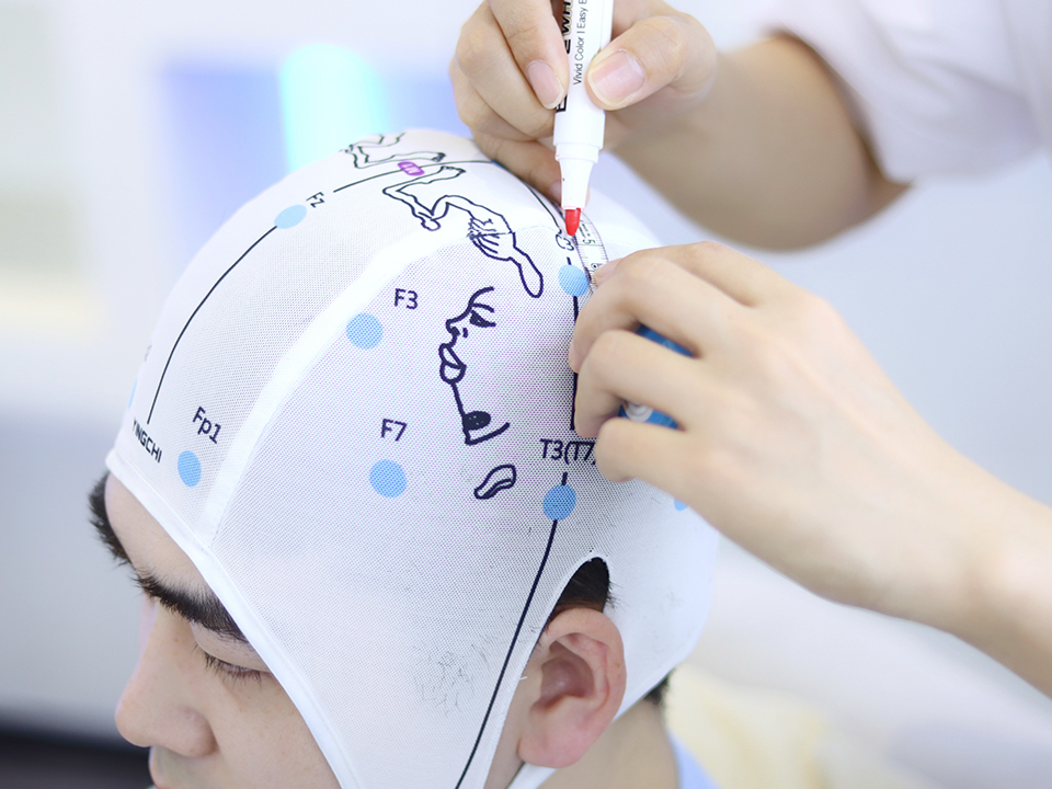
While modern rTMS clinics may use MRI-based neuronavigation, many rely on a more accessible, cost-effective method: the International 10–20 system. This scalp-based coordinate system, originally developed for EEG, offers a reproducible way to target specific cortical regions without imaging.
Why Use the 10–20 System in rTMS?
-
Standardization → Ensures that treatment sites are consistent between sessions and across clinics.
-
Accessibility → No need for MRI or neuronavigation hardware.
-
Efficiency → Target identification can be done in minutes with a tape measure.
From EEG Map to Stimulation Target
In rTMS, the 10–20 system helps locate cortical areas implicated in the condition being treated.
Key examples:
-
F3 (Left Dorsolateral Prefrontal Cortex – DLPFC)
→ Primary target in major depressive disorder.
→ Associated with emotion regulation, cognitive control. -
F4 (Right DLPFC)
→ Often targeted for anxiety, PTSD, or as part of bilateral depression protocols. -
SMA (Near FCz)
→ Used in obsessive–compulsive disorder protocols. -
M1 (C3/C4)
→ Primary motor cortex — used in motor threshold determination and post-stroke rehabilitation.
The Beam F3 Method
One of the most widely used adaptations of the 10–20 system for rTMS targeting is the Beam F3 method (Beam et al., 2009).
Steps (simplified):
-
Measure nasion–inion and left–right preauricular distances.
-
Identify Cz (intersection of these lines).
-
Use a set of proportional calculations to locate F3:
-
0.3 × nasion–inion distance along the midline from Cz toward nasion
-
0.6 × preauricular distance laterally from the midline point
-
Why F3 works:
MRI studies show that F3, as defined by this method, reliably corresponds to the left DLPFC in most people.
Advantages and Limitations vs Neuronavigation
Advantages:
-
No MRI required
-
Fast and inexpensive
-
Widely validated in depression treatment studies
Limitations:
-
Assumes average head–brain anatomy
-
Inter-individual variability can be significant (up to 1–2 cm difference in cortical target)
-
Cannot account for cortical folding patterns or lesions
Bottom line:
If precision is critical (e.g., post-surgical anatomy, research trials), MRI-based neuronavigation is preferred. For routine clinical use, the 10–20-based approach remains highly practical.
Clinical Examples
1. Depression
-
Protocol: Left DLPFC (F3) – High-frequency (10 Hz), 3,000 pulses/day, 4–6 weeks.
-
Rationale: Hypoactivity in left DLPFC in depression; stimulation restores prefrontal–limbic balance.
2. OCD
-
Protocol: SMA or orbitofrontal regions – Low-frequency (1 Hz) to reduce hyperactivity.
-
Landmark: SMA ~2 cm anterior to Cz on the midline.
3. Stroke Rehabilitation
-
Protocol: Contralesional M1 – Low-frequency inhibition to reduce interhemispheric imbalance.
-
Landmark: C3 or C4 depending on affected side.
When to Upgrade to Newer Methods
-
Research trials requiring high anatomical fidelity → Use MRI neuronavigation.
-
Unusual skull shapes or prior surgery → Imaging guidance avoids misplacement.
-
Multi-site protocols → High-density 10–10 or 10–5 systems allow finer targeting.
Closing Thoughts
The 10–20 system gives rTMS providers a low-cost, reliable method to find stimulation sites that have been validated in thousands of clinical studies.
While MRI-based navigation is more precise, the 10–20 approach remains a workhorse in both academic and private rTMS settings.
Mastering it ensures that even without expensive equipment, you can deliver effective, reproducible, and evidence-based brain stimulation.