Linking the 10–20 System to Brodmann Areas: From Scalp Landmarks to Cortical Function
 Introduction
The International 10–20 system gives us a reliable way to locate points on the scalp for EEG, rTMS, and other brain mapping techniques.
But clinicians and neuroscientists often need more than scalp coordinates — they need to understand what cortical regions lie underneath.
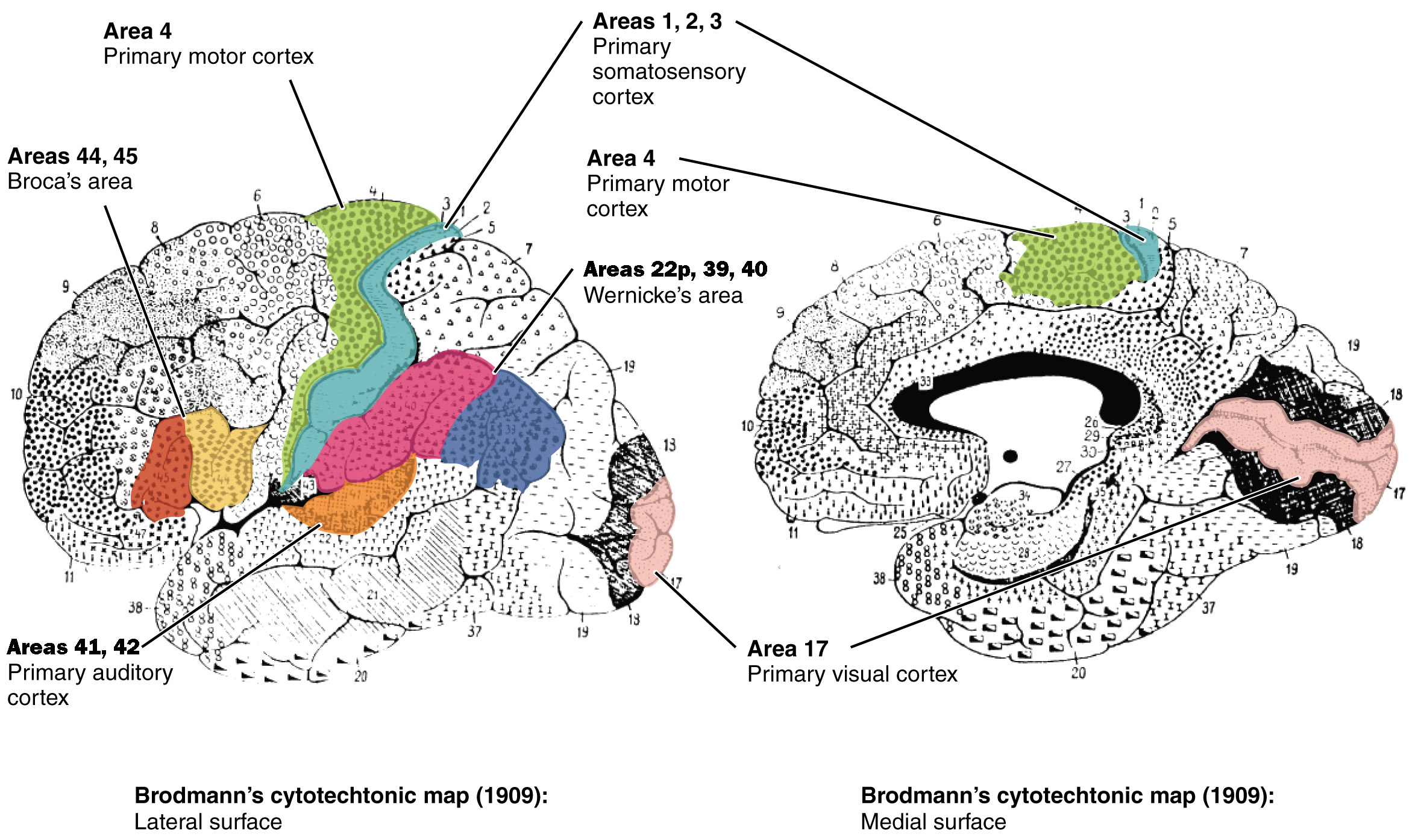
This is where Brodmann areas (BAs) come in. Developed by Korbinian Brodmann in the early 20th century, this cytoarchitectonic map divides the cortex into numbered areas based on cell structure.
By linking 10–20 positions to BAs, we can translate scalp landmarks into functional brain regions.
Why This Mapping Matters
-
rTMS → Helps target functionally relevant brain areas (e.g., BA46 in depression).
-
EEG → Interpreting focal abnormalities (e.g., BA17 in occipital seizures).
-
Neuropsychology → Relating cognitive deficits to electrode positions.
-
Research → Linking behavioural tasks to underlying cortical networks.
Mapping Principles
-
The 10–20 system gives surface landmarks, not exact cortical points.
-
Underlying BAs are approximate due to:
-
Skull shape variability
-
Cortical folding patterns
-
Individual brain size differences
-
-
MRI-based mapping offers the most precise localisation, but scalp–BA correlations remain valuable in practice.
Approximate Correspondence Table
| 10–20 Position | Approx. Cortical Location | Brodmann Areas | Functional Relevance |
|---|---|---|---|
| Fp1, Fp2 | Superior & middle frontal gyrus (frontal pole) | BA10 | Strategic planning, social cognition |
| F3 | Left dorsolateral prefrontal cortex | BA9, BA46 | Working memory, executive control |
| F4 | Right dorsolateral prefrontal cortex | BA9, BA46 | Attention, cognitive flexibility |
| F7 | Left inferior frontal gyrus (Broca’s area) | BA44, BA45 | Language production |
| F8 | Right inferior frontal gyrus | BA44, BA45 | Response inhibition, emotional processing |
| C3 | Left precentral gyrus (primary motor) | BA4 | Voluntary movement (contralateral) |
| C4 | Right precentral gyrus | BA4 | Voluntary movement (contralateral) |
| Cz | Midline pre/postcentral gyri | BA4, BA3/1/2 | Motor and somatosensory integration |
| P3 | Left superior parietal lobule | BA7 | Visuospatial processing |
| P4 | Right superior parietal lobule | BA7 | Spatial attention |
| O1, O2 | Primary visual cortex | BA17 | Visual perception |
| Oz | Midline occipital pole | BA17 | Visual midline integration |
| T3/T7 | Superior temporal gyrus | BA22 | Auditory processing (Wernicke’s on left) |
| T4/T8 | Superior temporal gyrus | BA22 | Auditory processing, social cognition |
| T5/P7 | Middle temporal/occipital junction | BA37, BA19 | Object recognition, reading |
| T6/P8 | Middle temporal/occipital junction | BA37, BA19 | Visual association, face recognition |
Clinical Examples
-
Depression → rTMS targets F3 (BA46) to improve prefrontal–limbic regulation.
-
Aphasia → EEG may show abnormalities over F7 (BA44/45) in left hemisphere lesions.
-
Visual seizures → Discharges at O1/O2 point toward BA17 involvement.
-
Neglect syndrome → Lesions affecting P4 (BA7) can cause spatial neglect.
Limitations and Cautions
-
BA mapping from scalp positions is approximate — always confirm with imaging in research or surgical planning.
-
Lateral displacement of gyri varies across individuals.
-
Some functional networks span multiple BAs (e.g., DLPFC includes BA9, 46, and parts of 8).
Closing Thoughts
Understanding how 10–20 positions relate to Brodmann areas bridges the gap between surface measurements and cortical function.
For clinicians, it sharpens diagnostic localisation and improves neuromodulation targeting.
For researchers, it strengthens the link between behavioural data and neural substrates.
While MRI and neuronavigation now offer precise cortical mapping, the 10–20–BA correlation remains a powerful clinical shortcut — especially in settings where imaging isn’t available.