The 10–20 System in EEG: Mapping Brainwaves with Precision
 Introduction
Electroencephalography (EEG) remains one of the most widely used and cost-effective tools for studying brain activity in both clinical and research settings.
From diagnosing epilepsy to monitoring sleep, EEG’s accuracy depends not just on the quality of the recording equipment but also on where the electrodes are placed.
That’s where the 10–20 system comes in — a standardized method for electrode placement that ensures reproducibility, comparability, and anatomical relevance.
While the system was originally designed for EEG, its principles are now applied in brain stimulation, neurofeedback, and even brain–computer interfaces. In EEG, however, its importance is unmatched.
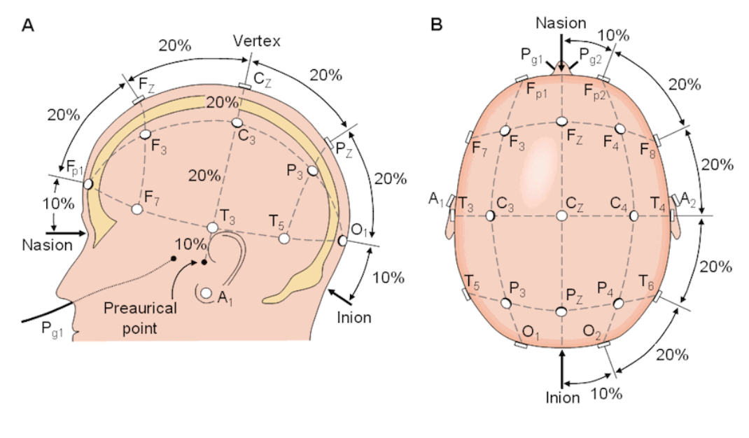
The Basics of EEG and the 10–20 System
EEG records electrical activity generated by cortical neurons, captured through electrodes placed on the scalp.
The 10–20 system defines the exact points for these electrodes based on proportional distances between anatomical landmarks (nasion, inion, and preauricular points), ensuring that each electrode is placed over a consistent cortical area across individuals.
This allows:
-
Reliable comparisons between recordings
-
Reproducible montages for clinical diagnosis
-
Standardized terminology across centres
Electrode Positions and Lobar Coverage
In standard 21-electrode clinical EEG:
-
Frontal pole (Fp1, Fp2) → Prefrontal cortex
-
Frontal (F3, F4, F7, F8) → Dorsolateral prefrontal, frontal eye fields, Broca’s area
-
Central (C3, C4, Cz) → Precentral (motor) and postcentral (sensory) gyri
-
Parietal (P3, P4) → Somatosensory association cortex
-
Occipital (O1, O2, Oz) → Primary visual cortex
-
Temporal (T3/T7, T4/T8, T5/P7, T6/P8) → Superior and middle temporal gyri, auditory cortex
Midline sites (Fpz, Fz, Cz, Pz, Oz) run along the sagittal midline.
Montage Types in EEG
The 10–20 system allows flexible electrode connection schemes called montages:
-
Referential Montage → Each electrode is compared to a common reference (e.g., mastoid, average of all electrodes).
-
Useful for detecting focal abnormalities.
-
-
Bipolar Montage → Electrodes are connected in chains, each channel representing the voltage difference between two adjacent electrodes.
-
Useful for localizing focal discharges and artifact differentiation.
-
-
Average Reference → Each electrode is compared to the average of all electrodes.
-
Useful in research and high-density EEG.
-
EEG Frequency Bands and Corresponding Sites
Electrode placement impacts how well you capture certain brain rhythms:
-
Delta (0.5–4 Hz) → Often dominant in frontal electrodes during deep sleep, diffuse in encephalopathy.
-
Theta (4–7 Hz) → Frontal midline theta in meditation; diffuse in drowsiness.
-
Alpha (8–13 Hz) → Best seen in occipital electrodes (O1, O2) when eyes are closed.
-
Beta (13–30 Hz) → Prominent in frontal/central regions, linked to active thinking and motor activity.
-
Gamma (>30 Hz) → Detected in research contexts; requires high sampling rates.
Clinical Applications
1. Epilepsy Diagnosis
-
Temporal lobe epilepsy → Spikes at T3/T7 or T4/T8
-
Frontal lobe epilepsy → Spikes at F3/F4 or F7/F8
-
Occipital lobe epilepsy → Spikes at O1/O2
Precise localization depends on electrode position accuracy.
2. Encephalopathy and Coma
-
Diffuse slowing, triphasic waves, or periodic discharges are best appreciated when the full 10–20 map is used.
3. Sleep Medicine
-
Sleep staging (NREM/REM) depends on occipital alpha attenuation, frontal slow waves, and central spindles — all identified through specific electrode sites.
4. Functional Brain Mapping
-
Pre-surgical mapping in tumour or epilepsy cases can use EEG reactivity in specific 10–20 positions.
Advantages of the 10–20 System in EEG
-
Universality → Same map used globally for diagnosis and research.
-
Reproducibility → Longitudinal patient follow-up uses identical positions.
-
Versatility → Works for routine, ambulatory, and high-density EEG.
Limitations in EEG Context
-
Spatial resolution is limited — standard clinical EEG may miss small foci.
-
Deep brain structures contribute minimally due to distance from scalp electrodes.
-
Inter-individual variation means the same position can overlie slightly different cortical gyri.
For greater precision, clinicians may use 10–10 or 10–5 extensions, adding more electrodes between standard positions.
Closing Thoughts
In EEG, the 10–20 system is not just a guideline — it is the foundation on which accurate diagnosis, meaningful research, and cross-centre collaboration are built.
Mastery of this system ensures that brainwave recordings are both technically sound and clinically interpretable.
Whether diagnosing epilepsy, staging sleep, or monitoring brain recovery, correct application of the 10–20 system can mean the difference between clarity and confusion in interpreting brain activity.