Beyond the 10–20 System: Newer Methods for Precision Brain Mapping
 Introduction
The International 10–20 system has served neuroscience, neurology, and psychiatry for over six decades, offering a standardized way to place EEG electrodes and target sites for brain stimulation.
But as technology and research have advanced, so have our mapping techniques.
Modern neuroscience demands greater precision, higher spatial resolution, and individualized targeting — especially in fields like epilepsy surgery, brain–computer interfaces (BCIs), and repetitive Transcranial Magnetic Stimulation (rTMS).
Enter the extended systems and imaging-based navigation.
Why Move Beyond the 10–20 System?
While the 10–20 layout is reliable and reproducible, it has limitations:
-
Spatial resolution: Only 21 standard positions; subtle abnormalities may be missed.
-
Inter-individual variability: A “C3” on the scalp may overly different gyri across people.
-
Precision needs: Advanced therapies require targeting within millimetres.
To address these, newer systems have added more electrodes or integrated imaging and computational models.
The 10–10 System
Overview
The 10–10 system doubles the number of electrodes by adding positions midway between those in the 10–20 layout.
Instead of just 10% and 20% intervals, this system uses 10% intervals only, creating about 64 electrode sites.
Advantages
-
Better spatial sampling → improved source localization.
-
Compatible with high-density EEG, tDCS, and research protocols.
-
Facilitates mapping of smaller cortical regions.
Applications
-
Epilepsy presurgical evaluation.
-
Advanced cognitive neuroscience experiments.
-
More precise rTMS targeting without neuronavigation.
The 10–5 System
Overview
The 10–5 system takes it even further, adding points at 5% intervals. This can yield 128–256 electrode sites.
Advantages
-
Very high spatial resolution → millimetre-level scalp coverage.
-
Allows detailed cortical mapping and improved modelling of current spread.
-
Integrates well with computational neuroimaging.
Applications
-
Brain–computer interface research.
-
Neural decoding experiments.
-
Precision neuromodulation studies.
High-Density EEG (HD-EEG)
What It Is
HD-EEG uses 64, 128, or 256 electrodes, often based on the 10–10 or 10–5 frameworks, to capture detailed spatial and temporal brain activity patterns.
Benefits
-
Better source reconstruction using advanced algorithms.
-
Improved detection of focal abnormalities.
-
Enhanced resolution for brain connectivity studies.
Challenges
-
More time-consuming setup.
-
Greater computational demands.
-
Higher equipment cost.
MRI-Based Neuronavigation
Overview
Instead of relying on scalp-based measurements, neuronavigation uses an individual’s MRI to directly map brain targets.
How It Works
-
MRI is uploaded to a neuronavigation system.
-
The target (e.g., left DLPFC, motor cortex hotspot) is identified in 3D space.
-
Infrared or electromagnetic tracking ensures precise coil or electrode placement.
Advantages
-
Millimetre precision.
-
Accounts for anatomical variations and lesions.
-
Gold standard for research and complex clinical cases.
Limitations
-
Requires MRI (added cost and patient exclusion in some cases).
-
Equipment cost is high.
-
More setup time per session.
Emerging Approaches
-
Connectome-Based Targeting
-
Uses individual structural and functional connectivity data (DTI, fMRI) to choose stimulation sites based on network patterns, not just anatomy.
-
-
Adaptive Closed-Loop Stimulation
-
Adjusts stimulation parameters in real-time based on EEG or other biomarkers.
-
-
Hybrid Mapping
-
Combines HD-EEG data with MRI for precision without full neuronavigation hardware.
-
Clinical Scenarios
-
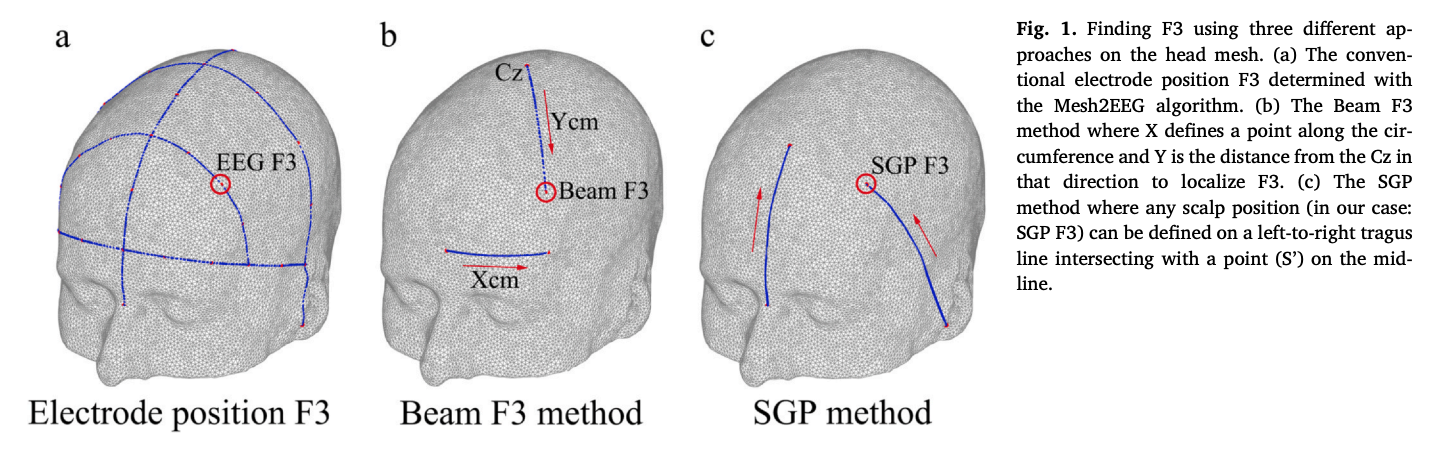
Routine EEG or rTMS in depression → Standard 10–20 or Beam F3 method is adequate.
-
Epilepsy surgery planning → 10–10 or HD-EEG improves localization.
-
BCI research → 10–5 or HD-EEG enables fine neural decoding.
-
Post-surgical or lesioned brains → MRI neuronavigation is essential.
Closing Thoughts
The 10–20 system remains a workhorse in brain mapping, but newer methods allow clinicians and researchers to push beyond its limits.
Whether you choose the 10–10 system, HD-EEG, or MRI-based neuronavigation depends on your goals, resources, and the precision required.
As brain science advances, the future is heading toward personalized, network-based mapping — where each patient’s unique anatomy and connectivity guide where and how we measure or stimulate the brain.