The 10–20 System: The Universal Map of the Human Brain
 Introduction
If you’ve ever stepped into an EEG lab or an rTMS clinic, you’ve probably heard someone mention positions like “F3”, “Cz”, or “Fp1.”
Far from being cryptic jargon, these labels come from the 10–20 system — a globally accepted method for placing electrodes or stimulation points on the scalp in a way that consistently relates to underlying brain regions.
Created by neurophysiologist Herbert Jasper in 1958, this system became the gold standard for electroencephalography (EEG) and has since been adapted for other fields, including brain stimulation therapies, neuroimaging, and cognitive neuroscience research.
Why It’s Called “10–20”
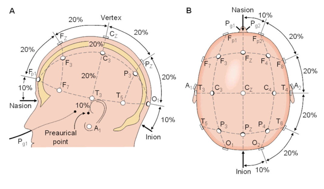
The name refers to the percentage distances between electrode sites:
-
Distances between fixed anatomical landmarks on the skull are measured.
-
Electrodes are placed at points that are 10% or 20% of those distances apart.
Because it’s based on proportional measurements, the 10–20 system works for any head size — from newborns to adults — and ensures that each point is positioned over roughly the same brain area in every individual.
The Anatomical Landmarks
Three reference points form the backbone of the system:
-
Nasion → The depression between the forehead and the nose bridge.
-
Inion → The bump at the back of the head (external occipital protuberance).
-
Preauricular Points → The small indentations just above the ear canals.
By measuring from front to back (nasion → inion) and side to side (left → right preauricular points), clinicians establish a mid-sagittal line and a coronal line. These intersect at Cz — the central reference point.
The Naming Code: Letters and Numbers
Every position is described by a letter (indicating brain region) and a number (indicating hemisphere):
Letters → Lobe/Region
-
Fp = Frontal pole
-
F = Frontal
-
C = Central (over motor strip, not a lobe)
-
P = Parietal
-
O = Occipital
-
T = Temporal
Numbers → Hemisphere
-
Odd numbers = Left side
-
Even numbers = Right side
-
z = Midline (“zero”)
Examples:
-
F3 = Left frontal lobe, lateral to midline.
-
C4 = Right central motor region.
-
Oz = Midline occipital area (visual cortex).
Mnemonics to Remember
A common way to remember the anterior-to-posterior order:
“Foolish People Catch Pigeons Occasionally”
-
Frontal pole → Frontal → Central → Parietal → Occipital
Or to walk around the head:
“Father’s Twin Can Play Tennis Outdoors”
-
Frontal → Temporal → Central → Parietal → Temporal → Occipital
The Standard EEG Layout
A typical 10–20 EEG setup uses 21 electrodes:
Midline: Fpz, Fz, Cz, Pz, Oz
Left hemisphere: Fp1, F3, C3, P3, O1, plus F7, T3, T5
Right hemisphere: Fp2, F4, C4, P4, O2, plus F8, T4, T6
This arrangement provides adequate coverage for:
-
Detecting seizure foci in epilepsy
-
Identifying encephalopathy patterns
-
Sleep staging and polysomnography
-
Baseline brain activity mapping
Why It Matters in Clinical Practice
The 10–20 system is not just a technical checklist — it’s a shared anatomical language used across multiple disciplines:
-
Neurology → Localising epileptic discharges, monitoring coma states.
-
Psychiatry → Targeting rTMS/tDCS for depression, OCD, or addiction.
-
Neuropsychology → Studying brain–behaviour relationships.
-
Research → Ensuring reproducibility across studies and subjects.
Because it’s standardized, an “F3” in one hospital is the same “F3” anywhere in the world.
Limitations and Modern Adaptations
While the 10–20 system is robust, it has limits:
-
Inter-individual variation → The same scalp point may overly different cortical gyri between people.
-
Resolution → 21 points are sufficient for EEG but too coarse for high-precision mapping.
-
Indirect mapping → It estimates cortical targets without direct imaging guidance.
These limitations have led to extended systems (10–10, 10–5) and MRI-guided neuronavigation for research and stimulation.
Closing Thoughts
The 10–20 system is one of neuroscience’s most enduring tools — as essential to brain mapping as latitude and longitude are to geography.
It is simple enough to learn in a few hours yet powerful enough to guide everything from epilepsy surgery planning to advanced brain stimulation protocols.
Understanding this system is a must for clinicians, researchers, and students who want to speak the universal language of brain coordinates.