Does Methylphenidate Change the Developing Brain? What a New 2026 Neuroimaging Review Reveals
Introduction: Beyond Symptom Control
Methylphenidate has been prescribed for attention-deficit/hyperactivity disorder (ADHD) for decades. Its effectiveness in reducing inattention, hyperactivity, and impulsivity is supported by substantial clinical evidence.
However, an important question remains:
When methylphenidate is administered during childhood, does it merely improve attention while the medication is active, or can it also influence the structural development of the brain?
This question has enormous implications for clinicians, parents, and individuals with ADHD.
Childhood and adolescence are periods of considerable neuroplasticity. Neural networks undergo structural reorganisation, synaptic pruning, changes in white matter organisation, and progressive maturation of cortical and subcortical regions.
A medication influencing dopamine and noradrenaline neurotransmission during these developmental periods could theoretically affect these processes.
But whether such effects are beneficial, harmful, temporary, or persistent requires careful investigation.
A systematic review published on 3 November 2026 in NeuroImage: Clinical by Liesbeth Reneman and colleagues offers new insights into this question.
The review, titled Methylphenidate-associated brain structural changes and neuroimaging predictors of treatment response in youth with ADHD, examines whether methylphenidate exposure is associated with measurable differences in brain structure and whether neuroimaging can help predict treatment response.
Its findings are intriguing, but they also highlight how much remains uncertain.
1. How Does Methylphenidate Influence the Brain?
Methylphenidate primarily inhibits dopamine and noradrenaline transporters, increasing the availability of these neurotransmitters at synapses.
These neurotransmitter systems are important for:
- Sustained attention and concentration
- Working memory and executive functioning
- Response inhibition and impulse control
- Motivation and reward processing
- Behavioural regulation
The therapeutic effects of methylphenidate involve several interacting brain networks, particularly frontostriatal circuits connecting the prefrontal cortex with the basal ganglia.
These networks support planning, behavioural inhibition, attention regulation, and goal-directed activity.
The distinction is between changes in brain function and changes in brain structure.
Functional changes occur when medication alters neurotransmission and neuronal activity. These may be observable within hours of administration.
Structural changes involve measurable differences in gray matter volume, cortical thickness, or white matter microstructure.
Such differences may reflect developmental processes, treatment-associated neuroplasticity, or pre-existing individual characteristics.
Importantly, an MRI difference does not automatically indicate either neurological improvement or damage.
2. What Did the 2026 Systematic Review Examine?
Reneman and colleagues searched MEDLINE and Embase for studies examining structural brain changes associated with methylphenidate treatment in children and adolescents with ADHD.
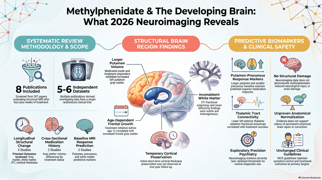
The researchers initially identified 507 publications, but only eight studies fulfilled their inclusion criteria.
These studies examined methylphenidate exposure lasting at least four weeks.
The eight publications were divided into three categories:
| Study category | Number |
|---|---|
| Longitudinal studies assessing structural change | 3 |
| Cross-sectional comparisons by medication history | 2 |
| Baseline MRI studies predicting treatment response | 3 |
Three publications were derived from the same randomized clinical trial, meaning the eight papers did not represent eight completely independent datasets.
The investigators examined three principal neuroimaging outcomes: gray matter volume, white matter integrity using diffusion tensor imaging (DTI), and cortical thickness.
An important qualification is that although the review discusses long-term effects, several included studies assessed treatment over only weeks or months. Therefore, the evidence is considerably stronger for identifying possible treatment-associated structural differences than for establishing permanent developmental effects.
3. Gray Matter Changes: The Putamen Emerges as an Important Region
One of the most interesting findings involves the putamen, a structure within the basal ganglia.
The putamen participates in motor regulation, reinforcement learning, habit formation, and aspects of action selection.
It forms part of the broader corticostriatal circuitry implicated in ADHD.
What did the studies find?
Villemonteix and colleagues (2015) compared children with ADHD who had received methylphenidate, never-medicated children with ADHD, and typically developing controls.
They reported differences in gray matter volumes involving several regions, particularly the putamen, insula, cerebellum, and parts of the frontal cortex.
Greater putamen volume was observed in the medicated ADHD group compared with the never-medicated ADHD group.
Separately, Chang and colleagues (2021) investigated structural MRI characteristics associated with subsequent medication response.
They found that individuals who responded well to methylphenidate had larger left putamen gray matter volumes and smaller bilateral precuneus volumes than poorer responders.
Together, these observations suggest that putamen structure may be associated with both medication history and clinical treatment response.
Does this mean methylphenidate normalises the ADHD brain?
Not necessarily.
Earlier neuroimaging studies have reported differences in basal ganglia volumes in ADHD, and some observational evidence suggests that stimulant exposure is associated with reduced differences between ADHD and typically developing groups.
However, structural normalisation is not equivalent to demonstrated functional improvement.
The putamen findings raise a hypothesis: methylphenidate may interact with developmental processes in circuits involved in attention, reinforcement, and behavioural control.
Yet, because the key putamen findings arose from cross-sectional and predictive analyses, they do not establish that methylphenidate itself caused the observed volume differences.
4. Frontal Cortical Development: Age May Matter
Another important finding concerns the frontal cortex.
The prefrontal cortex is crucial for executive functions such as planning, working memory, inhibitory control, and sustained attention.
Unlike many sensory and motor regions, parts of the prefrontal cortex continue maturing well into adolescence and early adulthood.
This prolonged developmental period may make frontal neural networks particularly responsive to environmental experiences and pharmacological exposure.
The significance of starting treatment before age 12
Chang and colleagues (2025) investigated associations between cumulative methylphenidate exposure and gray matter development.
The researchers found that greater cumulative exposure was associated with increased gray matter volume in several frontal cortical regions among participants who started medication before 12 years of age.
These associations were not observed among those who initiated treatment later.
Regions implicated included the superior and middle frontal cortices, orbitofrontal areas, and precentral regions.
This suggests a potentially important interaction between medication exposure and developmental timing.
However, the findings were not uniform across studies. Villemonteix and colleagues reported reduced middle frontal gray matter volume in medicated compared with never-medicated children with ADHD.
These differences demonstrate why the frontal cortical findings remain difficult to interpret.
Starting methylphenidate before 12 years of age should not be interpreted as a proven method for enhancing brain development.
The findings are observational and do not establish that earlier medication initiation produces better long-term neurological or functional outcomes.
They suggest that developmental stage may modify the association between methylphenidate exposure and brain structure.
5. White Matter: Could Methylphenidate Influence Neural Connectivity?
Gray matter contains neuronal cell bodies and much of the cortical processing machinery. White matter consists largely of axonal pathways connecting different brain regions.
Efficient white matter organisation is essential for communication between distributed neural networks.
In ADHD, connectivity between frontal regions, basal ganglia, and other cortical networks has been an important research focus.
Diffusion tensor imaging provides indirect measurements of white matter microstructure.
Two commonly used measures are:
Fractional anisotropy (FA): Describes the directional organisation of water diffusion, which is influenced by several tissue characteristics, including axonal organisation and myelination.
Mean diffusivity (MD): Measures the overall magnitude of water diffusion within brain tissue.
Changes in these parameters can suggest differences in tissue organisation, but neither measure directly establishes myelin growth or improved neural connectivity.
What did the review find?
Bouziane and colleagues (2019), using data from the randomized ePOD-MPH trial, identified age-dependent differences in white matter fractional anisotropy after methylphenidate treatment.
Voxel-based analyses demonstrated greater FA increases in some left-hemisphere association tracts and the corpus callosum among methylphenidate-treated boys.
Comparable changes were not observed in placebo-treated boys or adults.
However, the study did not find significant effects in its primary region-of-interest FA analyses.
Another study, by de Luis-García and colleagues (2015), reported lower mean diffusivity in several white matter tracts among medicated children with ADHD, depending on the analytical method used.
Other analyses yielded nonsignificant or different findings.
Overall, the review found that white matter results were subtle, heterogeneous, and inconsistent.
It is plausible that methylphenidate interacts with white matter maturation, particularly during childhood.
Nevertheless, the existing evidence cannot establish that methylphenidate improves myelination, strengthens connectivity, or permanently changes neural pathways.
6. Cortical Thickness: A Temporary Developmental Effect?
Perhaps the most intriguing observation concerns cortical thickness.
During typical brain development, cortical thickness changes through complex maturational processes.
A reduction in measured cortical thickness during adolescence is not necessarily pathological. It may reflect ordinary development involving synaptic, glial, and intracortical myelination changes.
Walhovd and colleagues (2020) examined cortical thickness following approximately 16 weeks of methylphenidate treatment in a randomized clinical trial.
They identified reduced cortical thinning, or relatively greater preservation of thickness, in a right medial cortical region among treated children compared with placebo.
The same effect was not observed in adults.
This suggested that methylphenidate might influence cortical developmental trajectories differently depending on age.
However, an important subsequent observation complicates the interpretation.
A four-year follow-up reported by van der Pal and colleagues (2024), discussed in the Reneman review as additional supporting literature, found no association between stimulant exposure and cortical thickness in the same region.
This raises the possibility that some structural effects observed after short-term treatment may not persist.
The evidence therefore suggests possible age-dependent modulation of cortical development, rather than a demonstrated permanent alteration in cortical anatomy.
7. Can MRI Predict Who Will Respond to Methylphenidate?
One of the most clinically relevant aspects of the review is its examination of neuroimaging predictors of treatment response.
Methylphenidate does not produce identical benefits in every person with ADHD.
Responses vary in symptom improvement, executive functioning, duration of benefit, and adverse effects.
The possibility of identifying likely responders using brain imaging is therefore attractive.
The review highlighted two principal findings.
Gray matter prediction: Chang and colleagues (2021) found that better responders had greater putamen volume and reduced precuneus volume.
White matter prediction: Chen and colleagues (2025) found that better responders had lower fractional anisotropy in the left anterior thalamic radiation.
The anterior thalamic radiation connects thalamic regions with frontal cortical networks and participates in cognitive and executive functioning.
These results support the possibility that differences in distributed brain networks contribute to variability in methylphenidate response.
However, these findings remain exploratory.
The studies do not establish a sufficiently validated imaging threshold, predictive accuracy, or reproducible clinical protocol for routine treatment selection.
MRI cannot presently determine with confidence whether an individual child will benefit from methylphenidate.
The findings represent a promising direction for precision psychiatry, not an established clinical test.
8. Does Methylphenidate Damage the Developing Brain?
This is understandably one of the most important concerns raised by parents.
The 2026 review does not demonstrate methylphenidate-induced structural brain damage.
Several studies instead identified associations with greater regional gray matter volume, while some reported changes in white matter organisation.
However, the absence of demonstrated structural damage in these studies should not be presented as definitive proof of long-term neurological safety.
The available research has important limitations.
First, the number of independent investigations remains small.
Second, MRI studies identify structural associations and cannot reliably determine whether these changes represent beneficial adaptation, altered developmental timing, or pre-existing characteristics.
Third, most studies had relatively short treatment or observation periods.
Fourth, many excluded children with psychiatric comorbidities, limiting generalisability to routine clinical populations.
Finally, the evidence cannot adequately characterise the consequences of continuous treatment extending across an entire childhood and adolescence.
Thus, two extreme interpretations are equally unsupported:
Methylphenidate permanently damages the developing brain.
Methylphenidate permanently repairs or normalises the ADHD brain.
Neither conclusion is justified by the available neuroimaging evidence.
9. Why This Review Requires Cautious Interpretation
Although most included publications received favourable methodological quality ratings, the authors themselves acknowledge several weaknesses.
Three of the eight publications came from a single randomized trial involving overlapping participants. Consequently, the effective number of independent datasets is closer to five or six.
Some studies included participants whose ages extended into adulthood despite the review’s emphasis on youth.
Several studies were predominantly male, and important psychiatric comorbidities were frequently excluded.
Treatment doses, exposure durations, imaging sequences, and analytical approaches varied substantially.
The review was not prospectively registered, and no formal advance review protocol was developed.
Importantly, the authors also noted that the quality-assessment approaches used did not comprehensively address all sources of bias. They recommended more rigorous risk-of-bias appraisal.
These limitations matter because statistical associations in neuroimaging studies may be sensitive to sample selection, analytical methodology, developmental stage, and clinical differences between groups.
Consequently, the review provides evidence of possible treatment-associated neurodevelopmental differences, rather than definitive proof of causal structural effects.
10. What Should Change in Clinical ADHD Practice?
The findings contribute to our understanding of ADHD neurobiology, but they do not currently justify major changes in prescribing practice.
Methylphenidate remains an evidence-supported pharmacological treatment for ADHD.
The NICE ADHD guideline recommends methylphenidate as a first-line medication option for children aged five years and over and young people when pharmacological treatment is clinically indicated, subject to applicable licensing and specialist prescribing requirements.
Treatment decisions should continue to be based on symptom severity, functional impairment, developmental needs, patient and family preferences, and the balance between potential benefit and adverse effects.
Clinical monitoring remains essential.
This includes attention to sleep, appetite, growth, weight, pulse, blood pressure, mood, and meaningful functional outcomes.
Structural MRI should not be routinely used to determine whether methylphenidate has improved brain development.
Neither should a larger putamen, altered cortical thickness, or particular DTI finding currently be regarded as a validated marker of therapeutic success.
Most importantly, treatment should aim to improve the child’s actual life: learning, relationships, emotional functioning, confidence, safety, and everyday independence.
An attractive neuroimaging finding has little clinical relevance unless it can eventually be connected to meaningful patient outcomes.
11. The Future: Towards Precision Treatment in ADHD
The emerging neuroimaging evidence raises an important possibility.
ADHD treatment response may depend not only on symptom presentation but also on underlying neural development and network organisation.
Future research may combine structural MRI, diffusion imaging, functional measures, neuropsychological assessment, and carefully characterised clinical outcomes.
Such approaches could help investigators identify developmental patterns associated with differential treatment response.
However, several questions need answering before these methods can become clinically useful.
Can findings be reproduced independently across hospitals and populations? Are MRI differences caused by medication or present before treatment? Do these structural differences predict improvement in daily functioning? Are effects sustained after discontinuation? Do children with comorbid autism, anxiety, learning disorders, or emotional dysregulation show similar patterns?
Answering these questions will require larger longitudinal studies, standardized imaging protocols, adequate representation of girls and diverse clinical populations, and follow-up extending well beyond the periods examined in most existing studies.
The ultimate objective should be clinically meaningful precision treatment rather than the identification of brain imaging abnormalities for their own sake.
Conclusion: A Medication That May Interact With Neurodevelopment
The 2026 systematic review by Reneman and colleagues adds an important perspective to the discussion of methylphenidate and the developing ADHD brain.
Its findings suggest that methylphenidate exposure is associated with measurable structural brain differences, particularly in subcortical regions such as the putamen.
Frontal cortical changes appear variable and potentially dependent on the developmental stage at which treatment begins.
White matter findings are inconsistent, while evidence concerning cortical thickness suggests possible short-term effects that may not persist.
Baseline neuroimaging characteristics also show preliminary associations with treatment response, opening possibilities for future research in precision psychiatry.
However, these findings do not establish that methylphenidate permanently changes brain structure, prevents abnormal brain development, or causes neurological harm.
The central lesson is that methylphenidate may interact with the developing brain in ways that extend beyond its immediate effects on attention, but the nature, clinical significance, and permanence of these interactions remain unresolved.
For clinicians, the priority remains evidence-based, individualised treatment and careful longitudinal monitoring.
For parents, the findings offer neither a reason for unnecessary alarm nor a justification for viewing medication as a method of correcting brain anatomy.
The most meaningful measure of successful ADHD treatment remains improvement in the individual’s functioning and quality of life.
Primary Reference
Reneman L, van der Pal Z, de Smidt D, Bipat S. (2026). Methylphenidate-associated brain structural changes and neuroimaging predictors of treatment response in youth with ADHD: a systematic review. NeuroImage: Clinical, 52, 104085.
DOI: https://doi.org/10.1016/j.nicl.2026.104085
Additional References
- Walhovd KB, et al. (2020). Methylphenidate effects on cortical thickness in children and adults with ADHD: A randomized clinical trial. American Journal of Neuroradiology.
- Bouziane C, et al. (2019). Methylphenidate-associated white matter microstructural findings from the ePOD-MPH trial.
- Villemonteix T, et al. (2015). Grey matter volumes in treatment-naïve versus chronically treated children with ADHD. European Neuropsychopharmacology.
- Chang and colleagues (2021, 2025). Studies of gray matter volume, medication response, and cumulative methylphenidate exposure, as reviewed by Reneman et al.
- van der Pal Z, et al. (2024). Stimulant medication use and apparent cortical thickness development in ADHD: A prospective longitudinal study.
- National Institute for Health and Care Excellence (NICE). Attention deficit hyperactivity disorder: diagnosis and management. NG87.
This article is an evidence-based interpretation of published research intended for educational purposes. It does not replace individualized clinical assessment or treatment planning.