Brain Mapping in Dementia and Memory Disorders: What qEEG Can Reveal About Alzheimer’s Disease and Cognitive Decline
 Can changes in memory be seen in the electrical activity of the brain?
Dementia is fundamentally a disorder of brain function. Long before severe memory loss develops, neurodegenerative diseases can alter the way neuronal networks communicate, synchronise and process information.
This has created growing interest in quantitative electroencephalography (qEEG), commonly called brain mapping, as a non-invasive method of studying brain function in people with:
- subjective memory complaints
- mild cognitive impairment (MCI)
- Alzheimer’s disease
- Lewy body dementia
- vascular cognitive impairment
- atypical cognitive decline
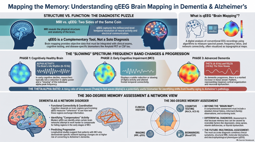
Unlike MRI, which primarily shows brain structure, qEEG measures the electrical dynamics of the functioning brain.
The most reproducible broad finding in Alzheimer’s disease is relatively intuitive:
As cognitive impairment progresses, brain electrical activity tends to become slower and less efficiently organised.
This may appear as:
↑ Delta and theta activity
↓ Alpha and beta activity
↓ Alpha peak frequency
Altered functional connectivity
Reduced network organisation
But brain mapping should not be confused with a definitive Alzheimer’s test.
qEEG does not diagnose dementia by itself. It provides complementary information about brain function that can be integrated with clinical examination, cognitive testing, imaging and disease-specific biomarkers.
What Is qEEG Brain Mapping?
A conventional EEG records tiny electrical signals generated by neuronal activity through electrodes placed on the scalp.
Quantitative EEG digitally analyses these recordings using mathematical algorithms.
Instead of relying exclusively on visual inspection of EEG waves, qEEG can measure:
- absolute spectral power
- relative spectral power
- delta activity
- theta activity
- alpha activity
- beta activity
- dominant or peak frequency
- hemispheric asymmetry
- coherence
- functional connectivity
- network organisation
- signal complexity
These measurements can then be represented as topographical brain maps.
The review by Popa and colleagues describes qEEG as digital EEG subjected to mathematical processing capable of examining frequency bands, signal complexity, connectivity and network characteristics. Importantly, the authors emphasise that qEEG should generally provide additional objective information alongside other diagnostic assessments rather than function as an immediate stand-alone diagnosis.
Why Is qEEG Particularly Interesting in Dementia?
Memory is not stored in one isolated location.
Successful cognition depends upon coordinated activity across distributed networks involved in:
- encoding information
- storing memories
- retrieving information
- attention
- language
- visuospatial processing
- executive functions
- orientation
- behavioural regulation
Neurodegenerative disorders progressively disrupt these networks.
EEG is therefore interesting because it measures neural activity with millisecond-level temporal resolution.
MRI may demonstrate:
“What does the brain look like?”
qEEG asks a different question:
“How is the brain functioning electrically?”
The two approaches are complementary.
The Classical qEEG Pattern in Alzheimer’s Disease
The most established electrophysiological feature of Alzheimer’s disease is EEG slowing.
The uploaded review describes common abnormalities including:
- alterations in delta and theta background activity
- reduced central alpha frequency
- reductions in alpha and beta activity
- decreased coherence between brain regions
A modern study replicated the major spectral pattern across two independent Alzheimer’s cohorts. Patients showed lower oscillatory alpha and beta power, resulting in a lower ratio of faster alpha/beta activity to slower delta/theta activity.
In simplified form:
Cognitively healthy brain
Relatively stronger organised alpha activity
↓
Early cognitive impairment
Subtle reduction or slowing of alpha activity
↓
Progressive neurodegeneration
Increasing theta activity
↓
More advanced dementia
Greater slow-wave activity, particularly theta and delta, with loss of faster organised rhythms
This progression should not be interpreted rigidly in every patient, but it captures one of the central electrophysiological observations in dementia research.
1. Alpha Activity: One of the Most Important Signals in Cognitive Decline
Alpha activity generally occupies approximately 8–13 Hz and is particularly prominent in posterior brain regions during relaxed wakefulness with the eyes closed.
In neurodegenerative disease, researchers have repeatedly observed:
- reduction in alpha power
- slowing of alpha frequency
- impaired alpha connectivity
- altered posterior alpha organisation
The review provided for this article reports reductions in alpha activity and a reduction in the central alpha frequency in Alzheimer’s disease.
This makes alpha particularly interesting in early cognitive impairment.
Rather than simply asking:
“How much alpha is present?”
modern qEEG can also examine:
Individual Alpha Peak Frequency
This is the dominant frequency within the alpha range.
A shift toward a slower peak frequency may indicate deterioration in the efficiency of large-scale neural processing.
2. Theta Activity and Dementia
Theta activity generally lies approximately between 4 and 8 Hz.
Increasing theta activity is one of the frequently reported findings as cognitive impairment progresses.
The uploaded review describes alterations in theta and delta activity as characteristic qEEG abnormalities in moderate and advanced Alzheimer’s disease.
Recent population-level clinical EEG research has also identified electrophysiological features related to cognition and Alzheimer’s pathology. In a 2024 study, data-driven EEG features correlated not only with cognitive test performance but also with PET metabolism and CSF Aβ42 measurements in the Alzheimer’s subgroup.
This is important because it suggests that EEG abnormalities are not merely random correlates of ageing—they may carry information related to the underlying neurodegenerative process.
3. Delta Activity: Increasing Cortical Slowing
Delta represents still slower electrical activity.
Marked delta activity during normal wakefulness generally becomes more prominent as cerebral dysfunction increases.
In dementia, excessive slow-wave activity may reflect:
- impaired cortical network organisation
- altered thalamocortical communication
- neuronal dysfunction
- progressive neurodegeneration
However, delta activity is not specific to dementia.
It may also occur with:
- delirium
- encephalopathy
- sedating medication
- metabolic disturbance
- structural neurological disease
- reduced alertness
This makes clinical context essential.
4. The Theta/Alpha Relationship
A potentially useful way of looking at dementia electrophysiology is not merely through individual frequency bands but through the balance between slower and faster rhythms.
As Alzheimer’s disease progresses:
Theta tends to increase
while
Alpha tends to decrease or slow
Consequently, ratios involving theta and alpha can shift.
Recent Alzheimer’s EEG research has found significant differences in spectral ratios such as theta/alpha and theta/beta between cognitively healthy individuals and patients with Alzheimer’s disease.
These measures may eventually become useful components of composite electrophysiological biomarkers.
But a theta/alpha ratio by itself should not be used to diagnose dementia.
5. Functional Connectivity: Dementia as a Network Disorder
Perhaps the most exciting development in modern dementia electrophysiology is the shift from studying isolated frequencies toward studying brain networks.
Cognition depends on communication between regions.
For memory to function normally, different areas of the brain must:
- exchange information
- synchronise appropriately
- communicate efficiently
- reorganise dynamically during cognitive demands
Neurodegenerative disease progressively disrupts this coordination.
qEEG can investigate these relationships through measures such as:
- coherence
- phase synchronisation
- amplitude-based connectivity
- network topology
The uploaded review describes reduced coherence in Alzheimer’s disease, including reductions involving theta, alpha and beta activity in frontal and central regions.
A 2024 study of mild cognitive impairment also demonstrated altered theta and alpha functional connectivity, including reduced frontal-temporal connectivity together with patterns interpreted by the authors as possible compensatory network activity.
This is a much more sophisticated model than simply saying:
“This patient has excess theta.”
The future is more likely to involve:
Which cognitive networks are failing—and which are compensating?
Mild Cognitive Impairment: Where qEEG May Become Most Useful
Mild Cognitive Impairment (MCI) occupies an important clinical space.
The person has measurable cognitive difficulty but remains substantially more independent than someone with established dementia.
The challenge is that MCI is heterogeneous.
Some people remain stable.
Some improve when reversible contributing factors are addressed.
Others progress toward:
- Alzheimer’s disease
- Lewy body dementia
- vascular dementia
- other neurodegenerative disorders
This makes MCI one of the most important areas for biomarker development.
Can EEG Predict Which Patients With MCI Will Develop Dementia?
Potentially—but this remains an evolving research area.
A 2024 study examining patients with MCI found that those who subsequently converted to dementia already demonstrated differences in EEG frequency characteristics, average connectivity and network topology before dementia became clinically established.
Even more recently, an 18-month longitudinal study published in 2025 evaluated EEG-based machine-learning approaches for identifying amnestic MCI patients progressing toward Alzheimer’s disease. The authors found that combining multiple EEG characteristics provided useful longitudinal discriminatory information.
This is where qEEG becomes particularly exciting.
The long-term goal is not simply to identify dementia after it is obvious.
The more valuable question is:
Can physiological changes identify high-risk cognitive decline earlier?
The answer from current research is promising—but not yet sufficiently validated for qEEG alone to determine an individual patient’s prognosis.
Subjective Memory Complaints Versus MCI
Many people attend memory clinics saying:
“My memory is getting worse.”
But subjective memory complaints do not automatically mean dementia.
Memory problems can result from:
- depression
- anxiety
- chronic stress
- poor sleep
- obstructive sleep apnoea
- ADHD
- medications
- alcohol
- metabolic disease
- thyroid dysfunction
- vitamin deficiencies
- normal ageing
Therefore, a brain map should never be the first or only answer to a memory complaint.
A structured assessment should first establish whether there is objective cognitive impairment.
Cognitive Testing and qEEG Answer Different Questions
This distinction is fundamental.
Cognitive testing asks:
How well is the person performing?
Tests may examine:
- memory
- attention
- language
- executive functioning
- visuospatial ability
- orientation
qEEG asks:
What electrophysiological patterns accompany that performance?
Clinical assessment asks:
Why is this happening?
The strongest approach therefore combines these different levels.
Why MoCA or ACE-III and qEEG Can Complement Each Other
A cognitive screening examination may identify deficits across domains such as:
- memory
- executive function
- visuospatial ability
- language
- attention
But two individuals with similar cognitive scores may have different underlying causes.
One may have early Alzheimer’s disease.
Another may have vascular cognitive impairment.
Another may primarily have depression-related cognitive symptoms.
Another may have a sleep disorder.
qEEG cannot independently resolve all of these diagnoses either.
But combining:
Clinical history + cognitive profile + electrophysiology + imaging/laboratory biomarkers
provides much more information than any individual test.
The uploaded review similarly notes a recommendation from earlier clinical neurophysiology literature to combine frequency analysis with cognitive assessment when evaluating dementia.
Can qEEG Diagnose Alzheimer’s Disease?
This requires an important distinction.
qEEG can detect functional abnormalities associated with Alzheimer’s disease.
But:
qEEG is not currently a disease-defining Alzheimer’s biomarker.
The 2024 revised Alzheimer’s Association criteria place disease-specific emphasis on biomarkers of Alzheimer pathology such as amyloid PET, CSF amyloid/tau measures and appropriately validated plasma biomarkers. EEG is not included among the core biomarkers capable of establishing the biological diagnosis of Alzheimer’s disease.
Therefore, a patient should not be told:
“Your brain map proves Alzheimer’s disease.”
A more accurate statement is:
“Your qEEG demonstrates a pattern of cerebral slowing or network dysfunction that may be compatible with cognitive impairment and should be interpreted alongside the clinical assessment and appropriate Alzheimer’s biomarkers.”
That distinction protects both scientific validity and clinical usefulness.
Brain Mapping Versus Alzheimer’s Blood Biomarkers
This distinction will become increasingly important.
Modern Alzheimer’s assessment is rapidly incorporating biological markers of disease pathology, particularly amyloid and phosphorylated tau measurements.
These biomarkers attempt to answer:
“Is Alzheimer-type pathology present?”
qEEG answers a different question:
“How is cerebral function currently organised?”
Therefore, the two approaches should not be considered competitors.
A future comprehensive memory assessment might combine:
Clinical cognitive phenotype
Structural MRI
Alzheimer’s blood/CSF/PET biomarkers where indicated
Functional electrophysiology
That would provide information about both:
Pathology and function.
Alzheimer’s Disease Versus Dementia
These terms are often incorrectly used interchangeably.
Dementia describes a syndrome of cognitive decline significant enough to impair functioning.
Alzheimer’s disease is one possible underlying neurodegenerative pathology.
Other causes include:
- Lewy body dementia
- vascular dementia
- frontotemporal dementia
- Parkinson’s disease dementia
- mixed dementia
- neurological and medical disorders
This distinction matters enormously when interpreting qEEG.
A brain map may identify cortical dysfunction without necessarily identifying its exact pathology.
Brain Mapping in Lewy Body Dementia
Lewy body disease is particularly interesting electrophysiologically.
Patients may develop:
- fluctuating cognition
- visual hallucinations
- parkinsonism
- REM sleep behaviour disorder
- attentional fluctuations
- visuospatial impairment
Because cognitive fluctuation is such a prominent feature, EEG abnormalities have been actively investigated as possible biomarkers.
Research criteria for mild cognitive impairment with Lewy bodies have identified quantitative EEG as a promising potential biomarker, although additional validation is still required.
Longitudinal research has also found EEG connectivity differences in MCI patients who later develop dementia, including patients progressing toward both Alzheimer’s and Lewy body dementia phenotypes.
This suggests that network analysis may eventually contribute to distinguishing neurodegenerative trajectories.
Brain Mapping in Frontotemporal Dementia
Frontotemporal dementia can present very differently from typical Alzheimer’s disease.
Early symptoms may involve:
- personality change
- disinhibition
- apathy
- loss of empathy
- language impairment
- executive dysfunction
rather than predominantly memory loss.
Because the underlying networks differ, researchers are investigating whether EEG connectivity and machine-learning approaches can help distinguish Alzheimer’s disease from frontotemporal dementia.
A 2025 study combining time-frequency characteristics and functional-connectivity measures demonstrated distinguishable network abnormalities between Alzheimer’s disease and frontotemporal dementia within its research dataset.
Again, this is promising research—not yet a stand-alone clinical diagnostic test.
Vascular Cognitive Impairment
Vascular brain disease can affect:
- processing speed
- executive function
- attention
- memory
- gait
- mood
The EEG consequences depend upon the location and extent of cerebrovascular pathology.
qEEG may reveal:
- slowing
- asymmetry
- regional abnormalities
- network disruption
But structural imaging such as MRI remains essential when vascular cognitive impairment is suspected.
Brain mapping should complement—not replace—appropriate neuroimaging.
Dementia Versus Delirium: An Important Clinical Distinction
Not every confused older adult has dementia.
Delirium typically involves an acute disturbance in attention and awareness that fluctuates over hours or days.
It can occur due to:
- infection
- medication
- electrolyte disturbance
- metabolic illness
- organ failure
- neurological disease
EEG frequently becomes diffusely slower during delirium and encephalopathy.
The uploaded qEEG review specifically notes that quantitative measures involving theta, delta and slow-frequency activity have been investigated in encephalopathy and in differentiating delirium.
This is another reason why:
Slow EEG activity does not automatically mean dementia.
Clinical context determines interpretation.
Can qEEG Measure Dementia Severity?
At a group level, increasing EEG slowing tends to correlate with worsening cognitive impairment.
But qEEG should not be used as a simple numerical dementia scale for an individual patient.
One cannot reliably say:
“This amount of theta means moderate dementia.”
Cognitive severity should still be assessed using:
- clinical functioning
- activities of daily living
- neuropsychological testing
- validated cognitive scales
- overall neurological and psychiatric assessment
qEEG can add physiological context to that evaluation.
Can Brain Mapping Track Dementia Over Time?
This is one of its most attractive potential applications.
Because EEG is:
- non-invasive
- repeatable
- relatively inexpensive compared with advanced imaging
- capable of directly measuring neural activity
serial recordings can potentially examine changes over time.
Researchers can investigate whether progression is accompanied by:
↓ Alpha frequency
↓ Alpha power
↑ Theta activity
↑ Delta activity
Altered connectivity
Changing network organisation
Resting-state EEG has already shown relationships with longitudinal cognitive performance and neurodegenerative outcomes in cohort research.
The future possibility is therefore a functional trajectory of cognitive decline, rather than a one-time colourful brain map.
What About EEG and Alzheimer’s Pathology Before Dementia?
One of the most fascinating questions is whether electrophysiological abnormalities appear before major clinical symptoms.
A 2024 study examined cognitively healthy individuals stratified according to CSF Alzheimer’s pathology and identified differences in functional connectivity in those with pathological amyloid/tau profiles, including patterns the researchers interpreted as possible compensatory connectivity.
This suggests that brain-network changes may begin before established dementia.
However, this should not be interpreted as justification for routine qEEG screening of healthy people for Alzheimer’s disease.
The 2024 Alzheimer’s diagnostic framework itself cautions against routine disease-biomarker testing in cognitively unimpaired individuals outside appropriate research contexts.
Brain Mapping and Machine Learning
Traditional EEG analysis evaluates predetermined characteristics such as:
- alpha power
- theta power
- delta activity
- coherence
Artificial intelligence changes the approach.
Instead of asking whether one particular variable is abnormal, machine-learning systems can analyse hundreds or thousands of electrophysiological features simultaneously.
Potential inputs include:
**spectral power
- frequency ratios
- connectivity
- entropy
- signal complexity
- network topology
- temporal characteristics**
Population-level EEG research has already demonstrated that data-driven approaches can extract features associated with cognitive performance, cerebral metabolism and Alzheimer’s biomarkers.
Longitudinal machine-learning studies are now attempting to predict progression from MCI toward Alzheimer’s dementia.
This is likely to be one of the most important directions for qEEG.
The Future May Be Multimodal, Not EEG Alone
The strongest future biomarker will probably not be:
EEG alone
or
MRI alone
or
a blood test alone.
Instead, precision dementia assessment may combine:
Clinical phenotype
What symptoms does the patient have?
Cognitive phenotype
Which domains are impaired?
Structural phenotype
What does MRI demonstrate?
Molecular phenotype
Are Alzheimer’s biomarkers abnormal?
Electrophysiological phenotype
How are neural oscillations and networks functioning?
Longitudinal trajectory
How are these measurements changing over time?
This moves memory assessment from simply asking:
“Is the MoCA low?”
toward:
“What is causing this patient’s cognitive impairment, what biological process is present, and how is brain function changing?”
What qEEG Cannot Do
Brain mapping should never be oversold.
At present, qEEG cannot independently:
- diagnose Alzheimer’s disease
- distinguish every type of dementia
- determine whether every memory complaint represents neurodegeneration
- accurately predict progression for every patient with MCI
- replace MRI
- replace detailed cognitive assessment
- replace Alzheimer’s disease-specific biomarkers
- determine dementia severity purely from map colours
The original review emphasises substantial biological and methodological variability, including effects of age, waking state, equipment, electrodes and artifacts. Its conclusion is therefore appropriately cautious: qEEG provides complementary objective information rather than an immediate diagnosis.
Who May Benefit From qEEG Brain Mapping?
Not everyone with occasional forgetfulness requires a brain map.
It becomes more clinically interesting in situations such as:
- mild cognitive impairment
- progressive memory complaints
- cognitive symptoms disproportionate to routine screening scores
- complex psychiatric versus neurological differential diagnosis
- unusual cognitive presentations
- possible Lewy body disease
- treatment or longitudinal monitoring
- research-oriented assessment
- need for broader objective cognitive characterisation
The test should answer a clinical question.
Technology without a clinical question simply produces data.
A 360-Degree Memory Assessment
An effective memory clinic should therefore go substantially beyond asking a few orientation questions.
A structured assessment may include:
1. Detailed history
- onset
- progression
- functional decline
- medications
- vascular risk
- sleep
- alcohol and substance use
- neurological symptoms
- psychiatric symptoms
2. Informant history
Family observations are often essential.
3. Domain-based cognitive assessment
Assessment may examine:
- attention
- memory
- language
- executive function
- visuospatial ability
- orientation
4. Screening instruments
Tools such as:
MoCA
ACE-III
may provide structured cognitive information.
5. Medical investigation
Depending on the presentation:
- laboratory evaluation
- structural neuroimaging
- neurological assessment
6. Disease-specific biomarkers
For selected patients, contemporary Alzheimer’s assessment may increasingly incorporate validated blood, CSF or PET biomarkers.
7. Functional brain assessment
qEEG can then provide another layer:
How is the brain functioning electrically?
Brain Mapping and Memory Assessment in Chennai
For patients and families concerned about memory loss, the most important first step is not obtaining a brain map—it is obtaining the correct diagnosis.
My approach to memory disorders is therefore built around comprehensive assessment rather than relying on any one investigation.
Depending on the clinical presentation, evaluation can integrate:
**Detailed geriatric psychiatric and neurological history
- Informant assessment
- MoCA / ACE-III and domain-wise cognitive evaluation
- Assessment of depression, sleep and medications
- qEEG / brain mapping where clinically useful
- Neuroimaging and laboratory investigations when indicated
- Integrated interpretation and longitudinal follow-up**
This is particularly valuable in patients with mild cognitive impairment or early cognitive symptoms, where establishing a baseline and following change over time may be more informative than waiting until dementia becomes obvious.
Frequently Asked Questions
Can qEEG diagnose Alzheimer’s disease?
Not independently. Alzheimer’s disease now has increasingly specific molecular biomarkers involving amyloid and tau. qEEG primarily provides information about brain function and network organisation rather than directly demonstrating Alzheimer pathology.
What does an Alzheimer’s brain map commonly show?
Common research findings include increased slow-frequency activity, reduced alpha and beta activity, slowing of alpha frequency and altered functional connectivity.
Does increased theta mean dementia?
No. Theta activity can increase for many reasons, including reduced alertness, medication effects and other neurological or metabolic conditions. It must be interpreted clinically.
Can qEEG detect mild cognitive impairment?
Research demonstrates measurable spectral and connectivity differences in some patients with MCI, but qEEG is not currently a stand-alone diagnostic test for MCI.
Can qEEG predict who will develop dementia?
Research is increasingly promising. Longitudinal studies have identified EEG connectivity and network differences among MCI patients who later progressed to dementia, and machine-learning models are being developed for progression prediction. These methods still require broader validation before being used as definitive individual prognostic tests.
Can qEEG distinguish Alzheimer’s from Lewy body dementia?
Possibly in the future. Distinct electrophysiological patterns have been reported, and quantitative EEG is being investigated as a biomarker for prodromal Lewy body disease, but it is not sufficiently definitive to replace clinical and established biomarker assessment.
Is qEEG the same as MRI?
No.
MRI → structure
qEEG → electrical function
The two investigations provide different and potentially complementary information.
Is brain mapping painful?
No. EEG is non-invasive. Electrodes record naturally occurring electrical signals from the scalp; they do not send electricity into the brain.
Memory Clinic and qEEG Brain Mapping in Chennai
Dr. Srinivas Rajkumar T
MBBS (Madurai Medical College), MD (AIIMS New Delhi), DNB, MBA (BITS Pilani)
Senior Consultant Psychiatrist
Apollo Clinic, Velachery, Chennai
Opposite Phoenix Marketcity
My clinical approach to dementia and memory disorders combines careful diagnosis, structured cognitive assessment and objective neurophysiological measurement where it provides meaningful additional information.
For patients with memory complaints, MCI or suspected dementia, qEEG should not be viewed as a shortcut to diagnosis.
Its greater potential lies in something more sophisticated:
**Cognitive testing tells us what functions are impaired.
MRI tells us about brain structure.
Molecular biomarkers tell us about disease pathology.
qEEG tells us how the brain is functioning electrically.**
Integrating these different layers may ultimately provide a much more complete understanding of cognitive decline than relying upon any single test.
Selected References
- Popa LL, Dragos H, Pantelemon C, Rosu OV, Strilciuc S. The Role of Quantitative EEG in the Diagnosis of Neuropsychiatric Disorders. J Med Life. 2020;13(1):8–15. doi:10.25122/jml-2019-0085.
- Jack CR Jr, Andrews JS, Beach TG, et al. Revised criteria for diagnosis and staging of Alzheimer’s disease: Alzheimer’s Association Workgroup. Alzheimer’s Dement. 2024. doi:10.1002/alz.13859.
- Kopčanová M, et al. Resting-state EEG signatures of Alzheimer’s disease are driven by periodic but not aperiodic changes. 2024.
- Li W, et al. Data-driven retrieval of population-level EEG features and associations with Alzheimer’s disease biomarkers. 2024.
- Hasoon J, et al. EEG functional connectivity differences predict future conversion from mild cognitive impairment to dementia. 2024.
- Ge Y, Yin J, Chen C, et al. An EEG-based framework for automated discrimination of conversion to Alzheimer’s disease in patients with amnestic mild cognitive impairment: an 18-month longitudinal study. Front Aging Neurosci. 2025;16:1470836. doi:10.3389/fnagi.2024.1470836.
- Jiang Y, et al. Altered EEG theta and alpha band functional connectivity in mild cognitive impairment. 2024.