Brain Mapping in Anxiety Disorders: What qEEG Can Reveal About the Anxious Brain
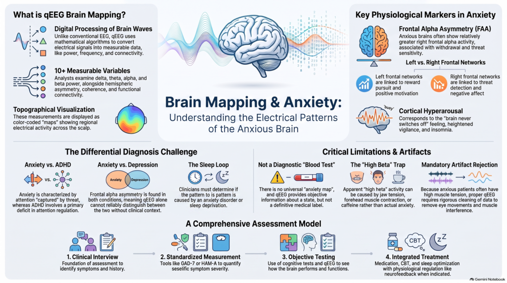 Can anxiety be seen in the electrical activity of the brain?
Anxiety disorders are diagnosed clinically. There is currently no EEG, qEEG brain map, MRI scan or blood test that can independently diagnose an anxiety disorder.
However, anxiety is accompanied by measurable changes in brain function. Research using quantitative electroencephalography (qEEG), commonly called brain mapping, has identified patterns involving frontal asymmetry, cortical arousal and regional electrical activity in people with anxiety disorders.
The important distinction is:
Brain mapping does not diagnose anxiety by itself. It can provide objective information about the neurophysiological state accompanying anxiety.
This is particularly relevant because two patients who both describe themselves as “anxious” may have very different underlying patterns—hyperarousal, panic, excessive worry, sleep disturbance, attentional dysregulation or depression-related anxiety.
What Is qEEG Brain Mapping?
A conventional EEG records electrical signals generated by neuronal activity using electrodes placed on the scalp.
Quantitative EEG (qEEG) mathematically processes these signals and converts them into measurable variables such as:
- delta power
- theta power
- alpha power
- beta power
- absolute and relative power
- dominant frequency
- hemispheric asymmetry
- coherence
- functional connectivity
- regional relationships between brain areas
These measurements can then be displayed as topographical brain maps.
The review by Popa and colleagues describes qEEG as digital EEG processed using mathematical algorithms to analyse frequency bands, signal complexity, connectivity and brain-network characteristics. Importantly, the authors emphasise that qEEG should generally provide additional objective information alongside clinical assessment rather than serve as an immediate diagnostic test.
Anxiety Is More Than “Too Much Thinking”
Anxiety involves coordinated changes across multiple systems.
A person experiencing significant anxiety may develop:
- excessive anticipation of threat
- hypervigilance
- autonomic arousal
- increased attention to danger
- difficulty disengaging from worry
- sleep disturbance
- muscle tension
- avoidance behaviour
- impaired concentration
- exaggerated startle
- panic symptoms
These experiences involve interactions between cortical and subcortical systems responsible for:
threat detection + emotional regulation + attention + autonomic control + behavioural inhibition
Therefore, researchers have investigated whether anxiety is accompanied by measurable changes in electrical brain activity.
Is There a Single qEEG Pattern for Anxiety?
No.
Just as there is no single clinical presentation of anxiety, there is no universal “anxiety brain map.”
Generalised anxiety disorder, panic disorder, social anxiety, obsessive worry and anxiety accompanying depression may produce overlapping but potentially different neurophysiological patterns.
The uploaded review highlights one of the most consistently discussed findings:
Frontal alpha asymmetry
The authors report that patients with anxiety have shown relatively greater right frontal alpha activity and that similar findings have been described in social phobia and panic disorder.
Understanding what this means requires understanding alpha activity.
Frontal Alpha Asymmetry in Anxiety
Alpha activity is commonly measured around 8–13 Hz.
A useful concept in EEG interpretation is that greater alpha power often corresponds to relatively reduced cortical activation in the underlying region.
Therefore, comparing alpha activity between the left and right frontal regions can provide information about relative frontal activation.
This is called:
Frontal Alpha Asymmetry — FAA
Research into emotional processing has proposed an approximate functional distinction:
Left frontal networks
More strongly associated with:
- approach behaviour
- reward pursuit
- behavioural activation
- positive motivation
Right frontal networks
More strongly associated with:
- withdrawal
- threat sensitivity
- avoidance
- negative affect
Anxiety is strongly associated with anticipatory threat and behavioural avoidance, making frontal asymmetry an attractive physiological marker to investigate.
The qEEG review reports greater right frontal alpha activity in anxiety and describes similar right-sided patterns in patients with social phobia and panic attacks.
But this should not be interpreted simplistically.
“Right-Sided Activity” Does Not Equal Anxiety Diagnosis
A brain map showing frontal asymmetry does not mean that anxiety has been definitively demonstrated.
Frontal alpha asymmetry may be influenced by:
- individual variability
- emotional state during recording
- depression
- sleep deprivation
- medications
- alertness
- recording conditions
- electrode characteristics
- muscle and eye-movement artifacts
The same review emphasises that EEG measurements can vary because of biological factors, waking state, equipment, electrodes and artifacts.
Therefore:
FAA is best interpreted as one physiological feature—not a diagnostic label.
Anxiety and Cortical Hyperarousal
Another important concept in anxiety is hyperarousal.
Many anxious patients describe:
“My brain never switches off.”
This subjective description can correspond to a broader physiological state involving:
- heightened vigilance
- increased sensory monitoring
- autonomic activation
- difficulty relaxing
- exaggerated response to threat
- insomnia
qEEG can help examine the balance between slower and faster electrical activity associated with different states of cortical activation.
For example, faster frequencies may become particularly interesting in patients who present with:
- restlessness
- marked cognitive arousal
- tension
- insomnia
- racing thoughts
However, an isolated increase in faster EEG activity cannot be labelled “anxiety,” because fast activity may also be influenced by muscle tension and other technical factors.
Beta Activity and Anxiety
Beta activity is broadly associated with alert cognitive processing.
In clinical qEEG interpretation, increased fast activity may sometimes be observed in people experiencing high arousal.
But interpreting beta requires caution.
Apparent beta activity can also be increased by:
- forehead muscle contraction
- jaw tension
- scalp muscle activity
- eye movements
- caffeine
- medications
- poor recording conditions
This matters particularly in anxiety, because anxious patients frequently have increased facial, forehead and jaw muscle tension.
Therefore:
An apparent “high beta” brain map should never automatically be interpreted as an anxious brain.
Artifact removal and clinical interpretation are essential.
Anxiety, Attention and Hypervigilance
Anxiety can significantly affect attention.
But the attentional difficulty in anxiety may be different from the attentional difficulty seen in ADHD.
Someone with anxiety may technically have the capacity to focus but repeatedly shift attention toward:
- perceived threat
- bodily sensations
- negative possibilities
- intrusive worries
- potential mistakes
- social judgement
This is sometimes better conceptualised as attention captured by threat rather than a primary deficit in attention regulation.
That distinction is clinically important.
A patient may say:
“I cannot concentrate.”
But the underlying reason could be:
ADHD
or
Anxiety
or
Depression
or
Sleep deprivation
or a combination of these.
This is why qEEG findings should be interpreted alongside the clinical history and, when appropriate, objective cognitive testing.
Brain Mapping Can Help Ask a Better Question
The wrong question is:
“Does this brain map prove anxiety?”
The more useful question is:
“What pattern of cortical activation, asymmetry and network function accompanies this patient’s anxiety symptoms?”
This shifts brain mapping from a binary diagnostic test toward neurophysiological phenotyping.
Generalised Anxiety Disorder
Generalised Anxiety Disorder (GAD) is characterised by persistent and excessive worry across multiple domains.
Patients may experience:
- chronic apprehension
- inability to control worry
- restlessness
- muscle tension
- fatigue
- irritability
- sleep disturbance
- impaired concentration
From a neurophysiological perspective, GAD is particularly interesting because it often involves persistent anticipatory processing rather than brief episodes of fear.
Brain mapping may therefore be used to examine broader patterns involving cortical arousal, frontal activity and connectivity.
However, the uploaded review does not identify a qEEG pattern sufficiently specific to diagnose GAD independently.
This is an important limitation.
Panic Disorder
Panic disorder represents a different form of anxiety.
Patients experience sudden episodes of intense fear accompanied by symptoms such as:
- palpitations
- chest discomfort
- breathlessness
- dizziness
- sweating
- tremulousness
- derealisation
- fear of losing control
- fear of dying
The qEEG review specifically notes that greater right frontal alpha activity has been reported in patients with panic attacks.
This is consistent with research interest in hemispheric asymmetry and threat-related emotional processing.
But again, this is an association rather than a diagnostic test.
Social Anxiety Disorder
Social anxiety involves excessive fear of:
- criticism
- embarrassment
- scrutiny
- negative evaluation
- rejection
The uploaded review reports greater right frontal alpha activity in social phobia as well.
This is particularly interesting because social anxiety strongly involves anticipatory avoidance.
A patient may experience marked physiological activation even before entering the feared social situation.
This creates potential opportunities for research using:
- resting qEEG
- task-related EEG
- anticipatory paradigms
- autonomic measurements
Anxiety Versus Depression on Brain Mapping
Anxiety and depression frequently coexist.
This can complicate qEEG interpretation.
Frontal alpha asymmetry has been investigated in both conditions.
The review notes that frontal alpha asymmetry is associated not only with depression but also with anxiety, and that parietotemporal asymmetry has been reported in both anxious and depressed subjects.
Therefore:
A frontal asymmetry pattern cannot reliably tell us whether the patient has anxiety or depression.
This is exactly why neurophysiological findings need clinical context.
Anxiety Versus ADHD
This is another increasingly important clinical differential diagnosis.
Both conditions can produce:
- distractibility
- procrastination
- restlessness
- impaired concentration
- forgetfulness
- difficulty completing tasks
But the mechanisms may differ.
ADHD
Attention regulation itself may be impaired.
Anxiety
Attention may repeatedly become captured by worry, threat or internal sensations.
The uploaded review describes research into increased theta and delta activity and altered theta/beta relationships in ADHD.
In contrast, the anxiety section particularly highlights frontal asymmetry.
These patterns cannot independently separate the disorders, but multimodal assessment may help clarify complex presentations.
Anxiety Versus Hyperarousal From Sleep Problems
Poor sleep can substantially alter EEG activity.
At the same time, poor sleep can produce:
- anxiety
- irritability
- reduced concentration
- emotional reactivity
- fatigue
- autonomic symptoms
This creates a common clinical loop:
Anxiety → poor sleep → increased arousal → worse concentration → more anxiety
Therefore, the interpretation of brain mapping in anxiety should always consider:
- sleep duration
- sleep quality
- insomnia
- circadian pattern
- daytime sleepiness
- possible sleep-disordered breathing
Otherwise, a physiological effect of sleep deprivation may be mistakenly attributed to an anxiety disorder.
Can qEEG Measure Anxiety Severity?
Not reliably by itself.
A more robust anxiety assessment combines:
Clinical interview
with
Standardised symptom measurement
such as:
- GAD-7
- HAM-A
- disorder-specific anxiety scales where appropriate
and, selectively:
Physiological or cognitive assessment
qEEG can contribute objective information, but symptom severity should not be inferred simply from how “abnormal” a colour map appears.
Can Brain Mapping Differentiate Different Anxiety Disorders?
At present, not reliably enough for routine stand-alone diagnosis.
Although research has reported physiological patterns in conditions such as:
- panic disorder
- social anxiety
- generalised anxiety
- depression with anxiety
the overlap between disorders is substantial.
This is partly because psychiatric diagnostic categories are based primarily on clinical syndromes, while EEG measures underlying physiological processes.
The same process—such as hyperarousal—may occur across several diagnostic categories.
This is precisely why qEEG may eventually become more useful for identifying biological dimensions than reproducing traditional diagnostic labels.
A Future Model: Anxiety Phenotypes Rather Than One “Anxiety Brain”
A useful future framework may involve identifying physiological subtypes.
For example:
Hyperarousal phenotype
Marked vigilance, insomnia and tension.
Threat-attention phenotype
Attention repeatedly captured by perceived danger.
Avoidance phenotype
Prominent behavioural withdrawal and frontal asymmetry.
Cognitive-worry phenotype
Persistent internal rumination and anticipatory processing.
Anxiety-depression phenotype
Combined motivational impairment and threat sensitivity.
These are conceptual examples rather than established diagnostic qEEG categories.
But they illustrate an important direction:
From diagnosis alone → toward individual neurophysiological phenotype.
qEEG and Neurofeedback for Anxiety
EEG becomes particularly relevant when discussing neurofeedback.
Neurofeedback attempts to provide individuals with real-time information about aspects of their physiological activity so that regulation can be trained.
qEEG may potentially contribute to:
- baseline electrophysiological assessment
- identification of target patterns
- protocol planning
- longitudinal monitoring
However, neurofeedback should not be presented as universally appropriate for every anxiety disorder.
Evidence strength varies according to:
- diagnosis
- protocol
- outcome studied
- equipment
- treatment design
It should therefore be integrated thoughtfully with established treatments rather than positioned as a replacement for them.
Brain Mapping Does Not Replace Evidence-Based Anxiety Treatment
Even when qEEG identifies an interesting physiological pattern, treatment remains guided primarily by clinical evidence.
Depending on diagnosis and severity, management may involve:
- psychoeducation
- cognitive behavioural therapy
- exposure-based therapy
- relaxation and arousal-regulation strategies
- sleep optimisation
- antidepressant medication
- other pharmacological strategies where appropriate
In disorders involving avoidance, particularly panic disorder and social anxiety disorder, exposure-based psychological treatment remains especially important.
Technology should strengthen treatment planning—not distract from interventions with established efficacy.
Can qEEG Predict Which Anxiety Medication Will Work?
At present, qEEG cannot reliably tell an individual patient:
“This is the anxiety medication that will definitely work for you.”
The uploaded review discusses qEEG primarily as an adjunctive clinical tool and emphasises limitations arising from methodological variability and inter-individual differences.
Medication selection therefore still depends on:
- specific anxiety diagnosis
- severity
- previous treatment
- comorbid depression
- bipolarity
- sleep
- substance use
- medical conditions
- side-effect profile
- patient preference
Why Artifact Control Is Especially Important in Anxiety
Anxious patients may unintentionally generate considerable EEG artifact.
Common sources include:
- jaw clenching
- forehead contraction
- frequent blinking
- eye movements
- muscle tension
- restlessness
These can contaminate the electrical recording, especially in faster-frequency ranges.
Therefore, proper qEEG requires:
good recording quality + artifact rejection + appropriate interpretation
A visually striking brain map generated from poor-quality EEG is still poor-quality data.
What qEEG Cannot Tell Us in Anxiety
qEEG currently cannot independently determine:
- whether someone definitely has an anxiety disorder
- which specific anxiety disorder is present
- whether anxiety is psychological or biological
- the exact severity of anxiety
- whether medication is required
- which medication will work
- whether psychotherapy will succeed
- whether symptoms are caused by anxiety rather than ADHD or depression
The correct role is adjunctive assessment.
Brain Mapping Versus MRI for Anxiety
These investigations provide different information.
MRI
Primarily evaluates brain anatomy and structure.
qEEG
Measures electrical brain activity and its temporal organisation.
Therefore, qEEG should be thought of as:
functional electrophysiological measurement
rather than a structural brain scan.
Can Brain Mapping Be Repeated After Treatment?
Yes.
EEG is non-invasive and can be repeated.
This creates potential applications for longitudinal assessment.
For example:
Before treatment
↓
clinical symptoms + baseline qEEG
↓
After treatment
↓
repeat symptoms + repeat physiological assessment
Researchers can then ask whether improvement in anxiety is accompanied by measurable changes in electrophysiology.
However, meaningful comparisons require similar recording conditions because EEG is influenced by multiple biological and technical variables. The review specifically warns that waking state, equipment, electrode factors and artifacts can alter qEEG measurements.
Who May Benefit Most From Brain Mapping?
Not every person with uncomplicated anxiety needs qEEG.
It may be more useful when there is:
- diagnostic uncertainty
- overlapping ADHD symptoms
- significant cognitive complaints
- anxiety associated with depression
- unusual treatment response
- treatment resistance
- prominent sleep or arousal problems
- consideration of neurofeedback
- need for objective longitudinal assessment
- research interest
The investigation should therefore answer a specific clinical question.
A More Objective Approach to Anxiety Assessment
The strongest model is not:
Brain mapping instead of psychiatric assessment
It is:
Clinical assessment + symptom measurement + cognition + neurophysiology
A comprehensive anxiety assessment can therefore include:
Detailed psychiatric interview
↓
Identification of the anxiety disorder
↓
Assessment of severity
↓
Evaluation for depression, ADHD, sleep disorders and substance use
↓
Objective cognitive or physiological assessment where clinically indicated
↓
qEEG / brain mapping in selected patients
↓
Integrated treatment plan
The Future of Brain Mapping in Anxiety
The future is unlikely to depend on a single EEG band.
Instead, increasingly sophisticated approaches may combine:
**spectral power
- asymmetry
- functional connectivity
- cognitive testing
- autonomic measures
- sleep data
- symptom scales
- machine learning**
This may eventually allow psychiatry to distinguish between patients who currently carry the same diagnostic label but have different underlying physiological patterns.
That is the larger promise of precision psychiatry.
Brain Mapping for Anxiety in Chennai
At my practice, the emphasis is on diagnosis-first psychiatry.
Technology is used when it helps answer a clinical question rather than as a substitute for psychiatric evaluation.
For selected patients with anxiety, assessment may incorporate:
**Comprehensive psychiatric evaluation
- Standardised anxiety measurement
- Screening for depression and ADHD
- Evaluation of sleep and substance use
- Cognitive testing when indicated
- qEEG / brain mapping
- Integrated treatment planning**
Management can then include appropriate combinations of:
- medication
- psychotherapy
- behavioural interventions
- sleep optimisation
- physiological self-regulation approaches
- neurofeedback where appropriately indicated
Frequently Asked Questions
Can qEEG diagnose anxiety?
No. Anxiety disorders remain clinical diagnoses. qEEG can provide complementary information regarding patterns such as frontal asymmetry and cortical arousal.
What brain-wave abnormality occurs in anxiety?
There is no single universal pattern. Research has particularly examined frontal alpha asymmetry. The uploaded qEEG review reports greater right frontal alpha activity in anxiety, including findings in social phobia and panic disorder.
Does high beta mean anxiety?
Not necessarily. Faster-frequency activity may be influenced by arousal, but muscle tension and other artifacts can also substantially increase apparent beta activity.
Can brain mapping distinguish anxiety from depression?
Not reliably on its own. Frontal and parietotemporal asymmetries have been reported in both conditions.
Can qEEG distinguish anxiety from ADHD?
Not independently. ADHD and anxiety can both impair concentration, but through different mechanisms. Clinical history, rating scales and objective cognitive testing may therefore be combined with qEEG where appropriate.
Is brain mapping painful?
No. EEG is non-invasive. Scalp electrodes record naturally occurring electrical activity; they do not send electricity into the brain.
Can qEEG choose an anxiety medication?
Not reliably. Medication selection continues to depend primarily on clinical diagnosis, previous treatment, comorbidity, safety and individual response.
Anxiety Assessment and Brain Mapping in Chennai
Dr. Srinivas Rajkumar T
MBBS (Madurai Medical College), MD (AIIMS New Delhi), DNB, MBA (BITS Pilani)
Senior Consultant Psychiatrist
Apollo Clinic, Velachery, Chennai
Opposite Phoenix Marketcity
My approach combines evidence-based clinical psychiatry with objective assessment where it provides meaningful additional information.
Brain mapping is not intended to replace the psychiatric interview.
It offers another window into the problem:
**Symptoms tell us what the patient experiences.
Cognitive testing tells us how the patient performs.
qEEG tells us something about the electrical activity occurring underneath.**
The future of anxiety assessment is likely to involve integrating all three rather than depending entirely on any single test.
Selected References
- Popa LL, Dragos H, Pantelemon C, Rosu OV, Strilciuc S. The Role of Quantitative EEG in the Diagnosis of Neuropsychiatric Disorders. J Med Life. 2020;13(1):8–15. doi:10.25122/jml-2019-0085.
- Heller W, Nitschke JB, Etienne MA, Miller GA. Patterns of regional brain activity differentiate types of anxiety. J Abnorm Psychol. 1997;106:376–385.
- Davidson RJ, Marshall JR, Tomarken AJ, Henriques JB. While a phobic waits: regional brain electrical and autonomic activity in social phobics during anticipation of public speaking. Biol Psychiatry. 2000;47:85–95.
- Wiedemann G, Pauli P, Dengler W, Lutzenberger W, Birbaumer N, Buchkremer G. Frontal brain asymmetry as a biological substrate of emotions in patients with panic disorders. Arch Gen Psychiatry. 1999;56:78–84.