Theta/Beta Ratio in ADHD: Why a High TBR Should Be Interpreted With Caution
 Quantitative EEG (QEEG) has attracted considerable interest in psychiatry because it offers an objective measure of brain electrical activity. Among the most discussed QEEG parameters in attention-deficit/hyperactivity disorder (ADHD) is the theta/beta ratio (TBR).
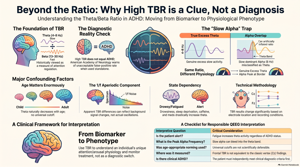
The concept appears simple. Theta represents relatively slow-frequency activity, commonly around 4–8 Hz, while beta represents faster activity, usually around 13–30 Hz. Dividing theta power by beta power gives the TBR. Historically, an elevated TBR was interpreted as reflecting cortical under-arousal or impaired attentional regulation.
However, contemporary EEG research suggests that the biology underlying this ratio is considerably more complex.
Why TBR Became Associated With ADHD
Early EEG studies frequently reported increased theta activity, reduced beta activity, and consequently elevated TBR in children with ADHD.
Initial meta-analyses found substantial differences between ADHD and control groups. These findings created enthusiasm that EEG might eventually provide an objective biological marker for ADHD.
Later studies, however, demonstrated much greater heterogeneity. Although some patients with ADHD clearly show increased TBR, many do not.
This distinction is fundamental:
A biological measure can differ between groups on average without being sufficiently accurate to diagnose an individual patient.
ADHD therefore remains primarily a clinical and developmental diagnosis, supported—not replaced—by objective testing.
A High TBR Does Not Automatically Mean ADHD
It is tempting to interpret a QEEG report containing an elevated theta/beta ratio as:
High TBR = ADHD.
That interpretation is too simplistic.
An elevated TBR should instead be regarded as an electrophysiological observation requiring clinical interpretation.
Several factors can influence it:
- age
- alertness and drowsiness
- sleep deprivation
- medication
- anxiety and arousal
- depression
- individual alpha frequency
- electrode location
- eyes-open versus eyes-closed recording
- reference montage
- frequency-band definitions
- artifact removal and preprocessing.
Consequently, TBR should never be interpreted independently of the clinical phenotype.
The Slow-Alpha Problem
One of the most important developments in modern TBR interpretation concerns individual alpha frequency.
Traditional QEEG systems usually divide the EEG spectrum into fixed bands:
Theta: approximately 4–8 Hz
Alpha: approximately 8–12 Hz
But the brain does not necessarily follow these mathematical boundaries.
Consider a person whose dominant alpha rhythm peaks around 8 Hz rather than 10 Hz.
Part of that individual’s alpha rhythm may extend below the conventional 8-Hz boundary. The software may therefore classify some physiologically meaningful slow alpha activity as theta.
The calculated theta power increases.
Because theta forms the numerator of the theta/beta ratio, the TBR may consequently appear elevated.
This means that two individuals can have essentially the same numerical TBR while having very different underlying electrophysiology.
Patient A
True excess activity around 5–7 Hz + relatively reduced beta.
Patient B
An alpha rhythm centred around 8 Hz spilling into the conventional theta range.
Both could produce a high TBR.
But their neurophysiology is not necessarily equivalent.
Therefore:
Peak alpha frequency should be examined before interpreting an elevated theta/beta ratio as pathological theta excess.
Recent research has increasingly distinguished a classical high-TBR phenotype from a slow-alpha-frequency phenotype, reinforcing the idea that ADHD itself may contain several electrophysiological subtypes.
TBR and the 1/f Problem
Modern EEG analysis has revealed another important complexity.
An EEG power spectrum contains not only identifiable oscillations such as alpha and theta but also an underlying background signal known as the aperiodic or 1/f component.
Traditional spectral analysis can mix the two together.
Therefore, what appears to be increased theta or decreased beta may sometimes partially reflect a change in the underlying aperiodic spectrum rather than a genuine alteration in a theta or beta oscillation.
Recent large-scale analyses have shown that conclusions regarding TBR can change depending on:
- the precise theta and beta frequency definitions,
- reference electrode,
- scalp location,
- recording state,
- individual alpha frequency,
- treatment of aperiodic activity,
- age correction,
- preprocessing methodology.
This is an important reminder that the TBR printed by software is not an immutable biological quantity.
It is partly dependent on how the EEG was recorded and analysed.
Age Matters
Brain electrical activity changes considerably with development.
Children naturally have more slow-frequency activity than adults. Theta generally decreases as the nervous system matures, while faster-frequency activity becomes more prominent.
Therefore, a TBR that might be relatively common in a child could carry a very different significance in an adult.
Statements such as:
“A TBR above 2.5 indicates ADHD”
are therefore scientifically problematic.
There is no universal TBR cutoff that can be applied across:
children, adolescents and adults; different electrodes; different recording conditions; and different analytical systems.
Age-appropriate normative interpretation is essential.
Alertness During Recording Matters
EEG is extremely state dependent.
Drowsiness increases slow-frequency activity.
A patient who slept poorly the previous night, is fatigued during testing, or gradually becomes drowsy during an eyes-closed recording may develop increased theta activity.
The resulting TBR can rise considerably.
Other relevant variables include:
- sleep duration the previous night
- caffeine
- recent exercise
- sedative medication
- psychotropic medication
- anxiety during recording
- time of day
- eyes-open versus eyes-closed condition.
This is why technically recording an EEG is only part of the process.
Knowing the state in which the brain was recorded is equally important.
Electrode Location Matters
There is no single universal “brain TBR.”
A TBR measured at Cz may differ significantly from one measured at Fz, frontal averages or posterior electrodes.
Much of the historical ADHD literature focused on central or vertex electrodes. Modern commercial QEEG reports, however, may generate theta/beta ratios across many scalp regions.
A mildly increased frontal TBR should therefore not automatically be interpreted as equivalent to the classical central TBR findings reported in older ADHD literature.
Interpretation should always specify:
where the ratio was measured, under what condition and using what reference values.
ADHD Is Neurophysiologically Heterogeneous
Perhaps the most important conceptual development is recognition that there may be no single ADHD EEG phenotype.
ADHD itself is heterogeneous.
One patient may predominantly experience:
- sustained-attention difficulty.
Another may primarily have:
- response-inhibition problems.
Another may perform extremely well when interested but struggle profoundly with:
- task initiation,
- organisation,
- procrastination,
- time perception,
- self-directed effort.
Others may have prominent emotional impulsivity or motivational dysregulation.
It is therefore unlikely that every individual meeting diagnostic criteria for ADHD will show the same resting EEG pattern.
Some may have elevated TBR.
Some may demonstrate slow alpha.
Some may have essentially normal TBR.
Others may show different spectral or connectivity abnormalities.
Thus:
Not having an elevated TBR does not exclude ADHD, and having an elevated TBR does not establish ADHD.
What About High-IQ Adults?
This becomes especially interesting in intellectually high-functioning adults.
A person with superior cognitive ability may compensate effectively for executive difficulties during school or during short structured cognitive tests.
They may perform well when:
- the task is interesting,
- feedback is immediate,
- testing is novel,
- environmental structure is high.
Their difficulties may emerge much more prominently when they need to independently:
- initiate assignments,
- organise multiple responsibilities,
- estimate time,
- maintain routine,
- switch tasks,
- tolerate boredom,
- persist without immediate reward.
Therefore, normal or near-normal performance on a brief attention test—or a relatively subtle QEEG pattern—does not automatically eliminate clinically significant executive dysfunction.
Objective tests need to be interpreted alongside real-world functional history.
Can TBR Predict Which Medication Will Work?
This is another area where caution is particularly important.
It is tempting to construct simple algorithms such as:
High TBR → stimulant
or
Low beta → atomoxetine
or
High beta → avoid stimulants.
The current evidence does not support such deterministic prescribing.
There are promising studies examining EEG predictors of medication response, including stimulants and atomoxetine. Some research suggests that changes occurring after medication exposure may ultimately prove more informative than the baseline EEG itself.
But no baseline TBR pattern is currently sufficiently validated to decide which psychiatric medication an individual patient should receive.
The more defensible model is:
Clinical diagnosis → rational medication selection → prospective measurement of response.
QEEG may then potentially contribute to response monitoring and physiological phenotyping.
QEEG May Be More Useful for Phenotyping Than Diagnosing
Perhaps the future of QEEG does not lie in asking:
“Does this brain map prove ADHD?”
A better question may be:
“What does this individual’s EEG tell us about attention, arousal and regulatory physiology?”
This shifts QEEG from being used as a diagnostic stamp toward becoming a physiological phenotyping tool.
Rather than focusing on TBR alone, a more comprehensive assessment can examine:
- absolute and relative theta
- alpha activity
- individual peak alpha frequency
- beta and high-beta activity
- regional distribution
- frontal asymmetry
- aperiodic activity
- task-related EEG
- connectivity where scientifically appropriate
- Continuous Performance Test findings.
These findings can then be combined with:
clinical interview + developmental history + psychodiagnostics + cognitive assessment + functional impairment.
This multimodal approach is more consistent with the direction of contemporary precision psychiatry.
QEEG and CPT Should Complement Each Other
A Continuous Performance Test (CPT) measures actual task performance rather than resting electrophysiology.
It may provide information regarding:
- omission errors
- commission errors
- reaction time
- reaction-time variability
- sustained attention
- response inhibition.
Interestingly, a patient may show high overall accuracy but nevertheless demonstrate increased commission errors or reaction-time variability.
That can indicate that the problem is not simply an inability to pay attention.
The difficulty may instead involve regulating attention and inhibition consistently across time.
This is precisely why combining clinical history, psychodiagnostics, CPT and QEEG can be considerably more informative than interpreting any single number.
A Practical Checklist Before Interpreting an Elevated TBR
When an elevated theta/beta ratio appears on a QEEG report, clinicians should ask:
- Was the patient fully alert throughout the recording?
- How well did the patient sleep the previous night?
- What is the individual peak alpha frequency?
- Could slow alpha be contributing to calculated theta?
- Where was TBR measured?
- Was it eyes open or eyes closed?
- Were age-appropriate normative values used?
- Could medication be influencing the EEG?
- Is depression, anxiety, fatigue or another condition contributing?
- Does the electrophysiology correspond with objective cognitive findings?
- Does the developmental and clinical history independently support ADHD?
The last question remains the most important.
How TBR Should Be Reported
Instead of writing:
“Elevated theta/beta ratio confirms ADHD.”
a more scientifically appropriate report would state:
“An elevated theta/beta ratio was observed, suggesting alteration in attention/arousal-related electrophysiological activity. TBR is influenced by age, alertness, individual alpha frequency, medication, recording methodology and other neuropsychiatric variables. It should therefore be interpreted alongside clinical assessment and objective cognitive measures and is not independently diagnostic of ADHD.”
Where the peak alpha frequency is slow, an additional qualification is useful:
“The relatively slow individual alpha frequency may contribute to apparent theta power because alpha activity may extend into the conventionally defined theta frequency range. The elevated TBR should therefore not automatically be interpreted as pathological theta excess.”
That distinction can substantially improve the scientific quality of QEEG interpretation.
The Future: From Brain Maps to Precision Psychiatry
Psychiatry needs objective biomarkers.
But the solution is unlikely to be one EEG ratio, one scan or one laboratory value.
Future precision psychiatry will probably combine multiple layers of information:
**clinical phenotype
- developmental history
- psychodiagnostics
- neuropsychological testing
- computerized cognitive assessment
- electrophysiology
- longitudinal treatment response.**
QEEG can potentially become an important part of this framework.
But its strength lies in adding information—not replacing clinical reasoning.
Conclusion
Theta/beta ratio remains an interesting electrophysiological measure, and elevated TBR may characterize a subgroup of individuals with ADHD.
However:
TBR is a physiological clue, not a diagnosis.
Before interpreting an elevated ratio, clinicians should examine individual alpha frequency, age, alertness, sleep, medications, recording condition, electrode location and the wider EEG spectrum.
Particular caution is required when peak alpha frequency is relatively slow, because slow alpha activity can enter the conventional theta band and artificially increase calculated TBR.
The future of QEEG in psychiatry therefore lies less in declaring:
“This brain map proves ADHD.”
and more in asking:
“What does this brain’s physiological pattern add to our understanding of this particular patient?”
That is a more cautious approach—but also a far more scientifically useful one.
About the Author & Clinical Assessment Services
Dr. Srinivas Rajkumar T is a Senior Consultant Psychiatrist with Apollo Hospitals, Chennai, with postgraduate training in Psychiatry from AIIMS New Delhi. His clinical interests include ADHD, cognitive assessment, QEEG, neurofeedback, interventional psychiatry and the responsible integration of emerging technologies into psychiatric practice.
At ATTN Clinic, Chennai, the focus is on a structured, multimodal assessment of attention and executive-function difficulties. Clinical diagnosis remains central, while selected patients may undergo additional computerized attention testing, psychodiagnostic assessment and exploratory QEEG brain mapping to develop a more detailed understanding of their cognitive and neurophysiological profile.
The aim is not to diagnose ADHD from a brain map, but to combine clinical expertise with objective measurements wherever these measurements can meaningfully improve assessment, treatment planning and longitudinal monitoring.
ATTN Clinic currently functions from:
Apollo Clinic, opposite Phoenix Market City, Velachery, Chennai
Appointments: +91 85951 55808
Email: srinivasaiims@gmail.com
Website: srinivasaiims.com
ATTN Clinic — Attention. Understood.