Ketamine and QEEG: What Brain-Wave Changes Are Seen After Ketamine Treatment?
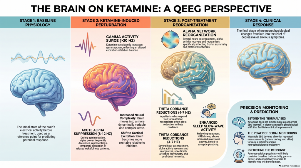 Ketamine is one of the fastest-acting treatments used in modern psychiatry, particularly for treatment-resistant depression and severe depressive symptoms. Because its effects can begin within hours, researchers have increasingly used Electroencephalography (EEG) and Quantitative EEG (QEEG) to study what happens to brain activity before, during and after ketamine treatment.
Research shows that ketamine does not produce one single “normalising” EEG pattern. Instead, it causes frequency-specific and region-specific changes in cortical activity.
Gamma activity increases after ketamine
One of the most consistently reported electrophysiological effects of ketamine is an increase in gamma-frequency activity, generally above 30 Hz.
Gamma oscillations are associated with local cortical processing and interactions between excitatory pyramidal neurons and inhibitory interneurons. Ketamine’s NMDA-receptor blockade alters this excitation-inhibition balance, producing measurable increases in gamma activity.
Several studies in depression have found increased gamma power following ketamine. However, greater gamma activity does not automatically mean greater antidepressant response. Baseline gamma activity and the magnitude of change may both influence the relationship with treatment response.
Alpha activity changes substantially
Alpha activity, usually between 8 and 12 Hz, is prominent during relaxed wakefulness, particularly when the eyes are closed.
During the acute ketamine state, alpha power frequently decreases. This has been demonstrated in EEG and Magnetoencephalography (MEG) studies.
Several hours after treatment, however, alpha activity may reorganise differently. Research using wearable prefrontal EEG in treatment-resistant depression has shown changes in:
- alpha power,
- frontal alpha asymmetry,
- alpha-related network activity.
This means that acute alpha suppression during ketamine administration and later alpha recovery or reorganisation are not contradictory. They represent different stages of the ketamine response.
Theta activity is highly regional
Theta activity generally occupies the 4-7 Hz range.
Ketamine studies have reported both increases and decreases in theta activity depending on:
- brain region,
- timing of the EEG,
- clinical diagnosis,
- treatment response,
- eyes-open or eyes-closed state.
Some studies have demonstrated increased frontal theta during acute ketamine exposure.
Other research has found that reduction in right-frontal theta activity may be associated with clinical improvement, particularly in treatment-resistant anxiety.
Baseline prefrontal theta activity has also been investigated as a potential predictor of ketamine response in treatment-resistant depression.
Therefore, simply stating that ketamine “increases” or “decreases” theta is inaccurate. Location and timing matter.
Theta cordance may be more informative than theta/beta ratio
Theta/Beta Ratio (TBR) is frequently discussed in QEEG, especially in ADHD assessment. However, it is not currently one of the strongest established ketamine biomarkers.
Ketamine research has focused more on:
- regional theta power,
- theta cordance,
- frontal theta activity,
- changes in theta after treatment.
Wearable EEG research in treatment-resistant depression has shown reductions in theta cordance among ketamine responders.
A falling theta/beta ratio alone should therefore not be interpreted as evidence that ketamine is working.
Beta activity also changes
Ketamine can alter beta activity, although findings vary depending on the beta frequency range.
Some studies have reported:
- reductions in low-beta activity,
- increases in higher-beta activity,
- redistribution of beta activity across cortical regions.
The overall pattern again supports the idea that ketamine produces reorganisation of cortical oscillations rather than a uniform shift toward faster brain waves.
Ketamine changes cortical excitation-inhibition balance
Modern EEG research is increasingly moving beyond conventional delta, theta, alpha, beta and gamma bands.
One promising measure is the aperiodic EEG exponent, which describes the background 1/f structure of the EEG spectrum.
Recent research in major depression has shown that ketamine can reduce the aperiodic exponent. This is consistent with a shift toward greater cortical excitation relative to inhibition.
Baseline aperiodic EEG characteristics may also contain information about subsequent antidepressant response.
This could become an important future direction in precision psychiatry and ketamine response prediction.
Ketamine increases EEG signal complexity
Ketamine also alters the complexity of brain activity.
Studies using measures such as:
- Lempel-Ziv Complexity,
- Multiscale Entropy,
have demonstrated increased neural complexity shortly after ketamine administration.
This suggests that ketamine temporarily moves cortical networks into a more dynamically variable state.
However, increased complexity does not necessarily predict antidepressant response. Some EEG changes may simply demonstrate that ketamine has altered brain physiology, rather than proving therapeutic benefit.
Sleep EEG also changes after ketamine
Ketamine’s neurophysiological effects continue after the acute treatment period.
Research in treatment-resistant depression has demonstrated increased slow-wave activity during subsequent Non-Rapid Eye Movement (NREM) sleep.
These changes have been associated with Brain-derived Neurotrophic Factor (BDNF), a molecule involved in synaptic plasticity.
Sleep EEG may therefore provide another window into the neuroplastic effects of ketamine.
Can QEEG predict ketamine response?
Research is promising, but there is currently no single validated QEEG biomarker that can reliably predict whether an individual patient will respond to ketamine.
Potential markers under investigation include:
- baseline frontal theta activity,
- theta cordance,
- gamma power,
- alpha activity and alpha asymmetry,
- EEG vigilance,
- signal complexity,
- aperiodic spectral characteristics,
- functional connectivity.
The future is likely to involve combining several EEG measures rather than relying on one brain-wave ratio.
Wearable EEG may make ketamine monitoring practical
Traditional research EEG systems may use 32, 64 or more electrodes and require specialised laboratory environments.
Portable EEG devices now make repeated measurements much easier.
This is particularly useful with ketamine because EEG can potentially be measured:
before treatment → during treatment → shortly after treatment → 24 hours later → during follow-up.
The real value of wearable EEG may therefore be serial within-patient monitoring, rather than a single QEEG brain map.
The most important principle
Ketamine does not simply make an abnormal EEG “normal.”
The better scientific model is:
baseline brain physiology → ketamine-induced perturbation → post-treatment network reorganisation → clinical response.
Current research suggests that changes in gamma activity, alpha regulation, regional theta activity, cortical complexity and excitation-inhibition balance may eventually help us understand why some patients respond rapidly to ketamine while others do not.
QEEG should therefore be viewed as an adjunctive neurophysiological tool, interpreted alongside diagnosis, symptoms, treatment history and clinical outcome.
Ketamine Treatment and QEEG Assessment in Chennai
Dr. Srinivas Rajkumar T, MD (AIIMS New Delhi), DNB, MBA (BITS Pilani) provides structured psychiatric assessment incorporating modern approaches including QEEG, objective cognitive assessment and evidence-based interventional psychiatry.
Consultations are available at Mind & Memory Clinic, Apollo Clinic Velachery, Chennai, opposite Phoenix Mall.
For patients being considered for ketamine or other advanced psychiatric treatments, the emphasis is on careful diagnosis, systematic treatment selection and objective monitoring wherever clinically useful.