Alexithymia and the Brain: What EEG and qEEG Reveal About Difficulty Understanding Emotions
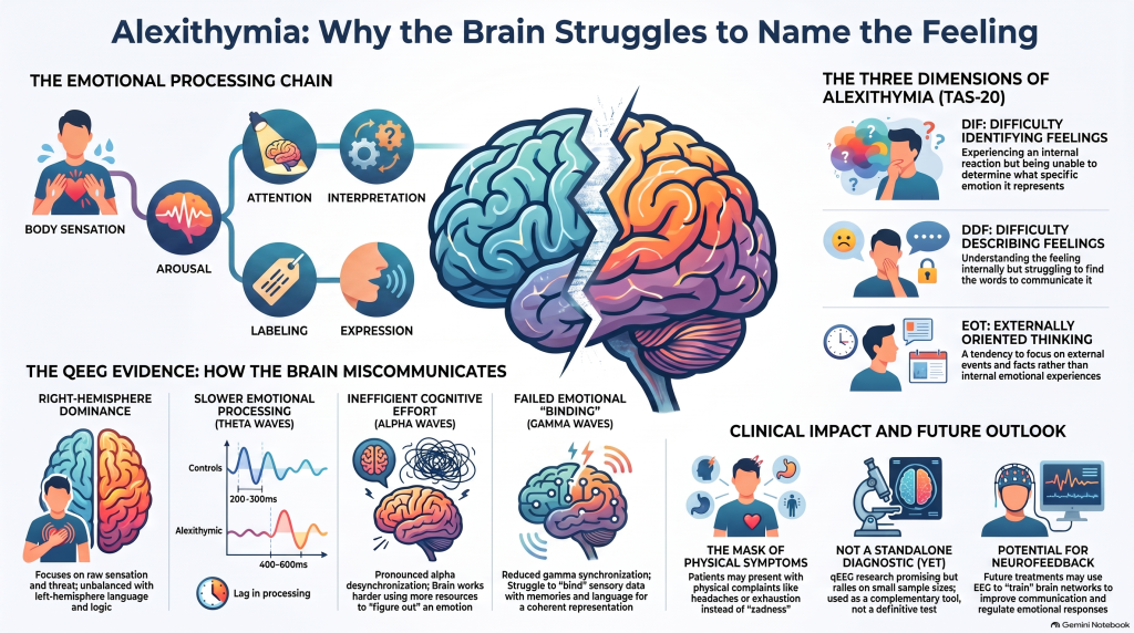 Some people experience emotions strongly but struggle to answer a seemingly simple question:
“What exactly am I feeling?”
They may notice a racing heart, chest tightness, irritability, restlessness or physical discomfort, yet find it difficult to identify whether the underlying emotion is fear, sadness, anger, embarrassment or something else.
This phenomenon is called alexithymia.
Alexithymia is not simply “having no emotions.” It refers to difficulties identifying and describing emotions, distinguishing emotions from bodily sensations, and a tendency toward externally focused rather than introspective thinking. Around 10% of the general population may have significant alexithymic traits, with substantially higher rates reported in conditions including autism, depression, schizophrenia, eating disorders, substance-use disorders and anxiety disorders.
A 2025 review in the Journal of Clinical Medicine examined an intriguing question:
Can we see aspects of alexithymia in the electrical activity of the brain?
The answer appears to be: possibly—but the science is still developing.
The review identified only eight eligible EEG studies, but several recurring patterns emerged involving brain asymmetry, connectivity and the alpha, theta and gamma frequency bands.
First, what is qEEG?
An electroencephalogram, or EEG, records electrical activity generated by populations of neurons using electrodes placed on the scalp.
Quantitative EEG, or qEEG, takes the EEG signal and mathematically analyses features such as:
- delta, theta, alpha, beta and gamma activity
- the relative distribution of these frequencies
- differences between brain regions
- asymmetry between the two hemispheres
- coherence or functional connectivity between regions
- topographic patterns of electrical activity
Rather than simply looking at an EEG tracing, qEEG attempts to quantify how different parts of the brain are functioning and communicating.
That becomes particularly interesting in alexithymia because emotional experience depends on several processes occurring together:
body sensation → emotional arousal → attention → interpretation → emotional labeling → verbal expression → regulation
Alexithymia may represent difficulty somewhere along this chain.
The central finding: alexithymia does not necessarily mean reduced emotion
One of the most interesting observations from these studies is that people with alexithymia often reported emotional experiences similar in intensity to people without alexithymia.
Show both groups an upsetting image or emotional film, and their subjective ratings may not differ dramatically.
Yet their EEG responses can be different.
This suggests an important distinction:
The problem in alexithymia may not be failure to experience emotion. It may be difficulty integrating, interpreting and labeling what is being experienced.
Across the reviewed studies, individuals with alexithymia frequently showed altered right-hemisphere activity, alpha and theta abnormalities, reduced connectivity between brain regions and weaker gamma synchronization.
1. Greater reliance on the right hemisphere
One of the strongest recurring observations was right-hemisphere dominance during emotional processing.
The right hemisphere has an important role in processing non-verbal emotional information, facial expressions, bodily signals and particularly negative or threat-related emotional cues.
Several EEG studies found greater right-sided involvement in people with alexithymia, particularly involving theta and alpha activity.
During emotional stimulation, for example, alexithymic participants showed greater alpha desynchronization over right centroparietal regions and increased theta synchronization, while control participants tended to show more balanced bilateral activation.
This does not simply mean that the “right brain is too active.”
A more useful interpretation is that emotional information may become disproportionately processed by networks involved in sensation and emotional arousal without adequate integration with systems involved in language, interpretation and cognitive regulation.
In other words:
The emotion may be generated—but converting that experience into “I am anxious,” “I am disappointed,” or “I feel rejected” may be more difficult.
2. The two hemispheres may communicate less efficiently
Emotion is not produced by one isolated brain region.
Recognising an emotion requires information to travel between networks dealing with:
- bodily sensations
- attention
- memory
- language
- autobiographical meaning
- executive control
- social understanding
Several studies found reduced interhemispheric coherence in alexithymia.
Reduced coherence was reported between frontal and central regions, as well as between the right frontal cortex and the left hemisphere. Altered connectivity was also found within default-mode-network regions involving areas such as the anterior and posterior cingulate cortex.
This provides an attractive neurological model for alexithymia.
The right hemisphere may register emotional and bodily information, while left-hemisphere language networks help categorise and verbalise it.
If communication between these systems is inefficient, the result may be:
“Something is happening inside me, but I cannot put it into words.”
That is remarkably close to the subjective experience many people with alexithymic traits describe.
3. Theta activity: more effort, but possibly slower emotional processing
Theta rhythms are involved in attention, memory and cognitive-emotional processing.
Some of the reviewed studies found increased right-sided theta activity during emotional stimulation.
Even more interesting was the timing.
In one experiment, theta synchronization associated with emotional processing peaked around 400–600 milliseconds after the stimulus in alexithymic individuals, compared with approximately 200–300 milliseconds in controls.
This raises the possibility that emotional information is not completely absent—it may simply be processed differently or less efficiently.
The brain may need additional cognitive resources to interpret something that other people identify more automatically.
4. Alpha activity: emotional processing may require more cognitive effort
Alpha activity is frequently misunderstood as simply representing an “inactive brain.”
In reality, changes in alpha activity can reflect shifts in cortical inhibition, attention and cognitive workload.
Several alexithymia studies found pronounced alpha desynchronization, particularly over parietal and centroparietal regions during emotional stimulation.
Alpha desynchronization generally accompanies greater cortical engagement.
One proposed interpretation is therefore that people with alexithymia may actually be allocating more attentional resources to emotional information—but doing so inefficiently.
Instead of automatically understanding:
“She looks disappointed, and that makes me feel guilty,”
the brain may focus intensely on external perceptual information while struggling to connect those signals with an internal emotional category.
The review describes this possibility as a form of attentional hyperfocus with increased cognitive load.
5. Gamma activity: difficulty “binding” emotion into a meaningful whole
Gamma oscillations are particularly interesting because they are associated with the integration—or “binding”—of information distributed across multiple neural networks.
When most people view emotionally negative images, gamma activity and synchronization increase as sensory information becomes connected with:
- attention
- memory
- previous experiences
- meaning
- emotional appraisal
In one of the reviewed studies, people without alexithymia showed an increase in gamma power and phase synchronization approximately 400–450 milliseconds after negative emotional stimuli.
People with alexithymia did not show the same robust gamma response.
The authors suggest that reduced gamma synchronization could represent weaker integration between the sensory experience of an emotion and the memories, concepts and language required to understand it.
Put simply:
The individual may receive the pieces of the emotional experience but have more difficulty assembling those pieces into a coherent emotional representation.
A useful model: high arousal + poor labeling
Perhaps the most clinically useful interpretation of the research is that some forms of alexithymia may involve a mismatch between emotional arousal and emotional understanding.
Imagine the brain receiving strong bodily signals:
Heart beating faster.
Muscles tightening.
Stomach uncomfortable.
Attention narrowing.
Something feels wrong.
But the cognitive system cannot efficiently transform those signals into:
“I am anxious because tomorrow’s meeting feels threatening.”
Instead, the person may simply report:
“I don’t know. I just don’t feel good.”
The review proposes that alexithymia can involve heightened autonomic and right-hemisphere responses alongside weaker top-down regulation and emotional labeling.
This helps explain why alexithymia can sometimes present clinically through physical complaints, irritability, unexplained distress or difficulty communicating emotions rather than obvious sadness or anxiety.
Alexithymia is probably not one single brain pattern
Another important development is recognition that alexithymia itself is heterogeneous.
The widely used Toronto Alexithymia Scale examines three major dimensions:
DIF — Difficulty Identifying Feelings
A person experiences an internal reaction but struggles to determine what emotion it represents.
DDF — Difficulty Describing Feelings
The individual may understand the feeling internally but struggles to communicate it verbally.
EOT — Externally Oriented Thinking
Attention is directed predominantly toward external events rather than internal emotional experiences.
The review proposes that these dimensions may eventually correspond to somewhat different neural mechanisms.
For example, difficulty identifying feelings could be more strongly related to heightened autonomic arousal and right-hemisphere processing, whereas difficulty describing feelings might involve weaker integration with left-hemisphere language networks. Externally oriented thinking may involve disproportionate attention to external sensory information.
This raises an intriguing possibility:
There may eventually be several neurobiological subtypes of alexithymia rather than one universal “alexithymia brain.”
Why this matters in psychiatry
Alexithymia is particularly relevant because it appears across multiple psychiatric and neurological conditions.
Someone presenting with depression may not say:
“I feel sad and hopeless.”
Instead they may describe:
- exhaustion
- headaches
- body pain
- sleep disturbance
- irritability
- loss of motivation
- vague internal discomfort
Similarly, someone with anxiety may describe palpitations and gastrointestinal symptoms while having difficulty recognizing the underlying fear.
A person with autism may experience intense emotional arousal yet struggle to identify, communicate or regulate it.
Alexithymia can therefore complicate clinical assessment because our conventional psychiatric interview depends heavily on a person’s ability to describe their internal state.
Could qEEG provide a more objective window?
This is where qEEG becomes scientifically interesting.
Traditional assessment asks:
“How do you feel?”
EEG asks a different question:
“What is the brain doing while emotional information is being processed?”
The reviewed studies suggest that physiological differences can sometimes be present even when subjective emotional ratings appear similar.
In the future, objective measurements might potentially complement clinical interviews and psychological scales by examining:
- hemispheric asymmetry
- alpha and theta responses
- functional connectivity
- coherence
- gamma synchronization
- resting-state versus emotion-task responses
But there is an important qualification.
qEEG cannot currently diagnose alexithymia
The research is promising, but we should not convert interesting neuroscience into premature clinical certainty.
Only eight EEG studies met the criteria for this 2025 review, and many involved relatively small samples. Some studies included only 20–24 participants, and participants were frequently university students or other relatively homogeneous populations.
There was also considerable variation in:
- electrode numbers and placement
- EEG reference methods
- definitions of frequency bands
- preprocessing and artifact removal
- emotional tasks
- resting-state versus task-based recordings
- alexithymia scales and cutoff scores
The review itself was mechanistic rather than a full systematic review and did not include formal risk-of-bias or PICOS assessment.
The authors specifically highlight methodological inconsistencies and the need for standardized protocols and larger, more diverse samples.
Therefore:
qEEG should not currently be considered a standalone diagnostic test for alexithymia.
Its more realistic role is as an emerging research and complementary neurophysiological tool.
Could neurofeedback eventually help?
This is perhaps the most exciting—but also most preliminary—question.
If some alexithymic traits are associated with identifiable patterns of cortical activity and connectivity, could those networks eventually be trained?
The review proposes several possibilities.
For example, neurofeedback could theoretically target excessive or dysregulated right frontal or right parietal alpha/theta activity. Other interventions might attempt to improve communication between hemispheres.
EEG could also potentially be used longitudinally:
Baseline → psychological intervention → repeat EEG → examine whether neural changes accompany clinical improvement.
The authors discuss neurofeedback, mindfulness-based interventions, psychotherapy and neuromodulation as areas deserving further study. However, most existing EEG research is cross-sectional rather than treatment-based, so these possibilities should currently be regarded as research directions rather than established treatments for alexithymia.
The future may be multimodal
EEG has excellent temporal resolution—we can observe brain activity changing within milliseconds.
Its major limitation is spatial resolution.
Future research may therefore combine EEG or qEEG with:
- fMRI
- functional near-infrared spectroscopy (fNIRS)
- MEG
- heart-rate variability
- electrodermal activity
- autonomic measurements
- psychological and cognitive testing
The review specifically proposes combining EEG with modalities such as fMRI, PET, NIRS and MEG to better identify the networks underlying altered emotional processing.
This approach reflects a broader movement in psychiatry: moving beyond a single questionnaire or symptom description toward multimodal measurement of brain, behaviour and subjective experience.
The most important insight
The neuroscience of alexithymia challenges a common misconception.
A person who cannot describe their emotions may not be emotionally empty.
They may be experiencing considerable emotional and physiological activity while lacking an efficient mechanism for converting that activity into conscious, differentiated and verbally accessible feelings.
The emerging EEG picture is therefore not simply:
“Less emotion.”
It may instead involve:
more arousal, altered attention, inefficient integration and weaker emotional labeling.
Across the studies reviewed, right-hemisphere predominance, reduced communication between brain regions, altered alpha and theta activity and reduced gamma synchronization repeatedly appeared as potential neurophysiological correlates.
That distinction matters.
Because once emotional difficulty is understood as a problem of processing and integration rather than absence, it opens the door to better assessment—and potentially, in the future, more targeted treatment.
Clinical perspective
In my psychiatric practice, I am particularly interested in moving beyond symptom labels toward a more structured understanding of how attention, cognition, emotion and brain function interact. Clinical assessment remains the foundation of diagnosis, while tools such as cognitive testing, EEG/qEEG and emerging neurophysiological measures can potentially provide additional objective information when used appropriately.
Dr. Srinivas Rajkumar T
Senior Consultant Psychiatrist, Apollo, Chennai
MD Psychiatry — AIIMS New Delhi
ATTN Clinic — Attention. Understood.
Apollo Clinic, opposite Phoenix Market City, Velachery, Chennai
Appointments: +91 85951 55808
This article is educational and is based primarily on the 2025 mechanistic review “Neural Correlates of Alexithymia Based on Electroencephalogram (EEG)” by Chmiel and colleagues. EEG/qEEG findings in alexithymia remain an evolving research area and should not be interpreted as an established standalone diagnostic test.