Can qEEG Help Detect Dementia? What Brain-Wave Patterns and Machine Learning Are Revealing
 Dementia is traditionally diagnosed through clinical assessment, cognitive testing, functional evaluation, laboratory investigations and brain imaging. Increasingly, however, researchers are asking another question:
Can the electrical activity of the brain provide objective clues to early cognitive decline?
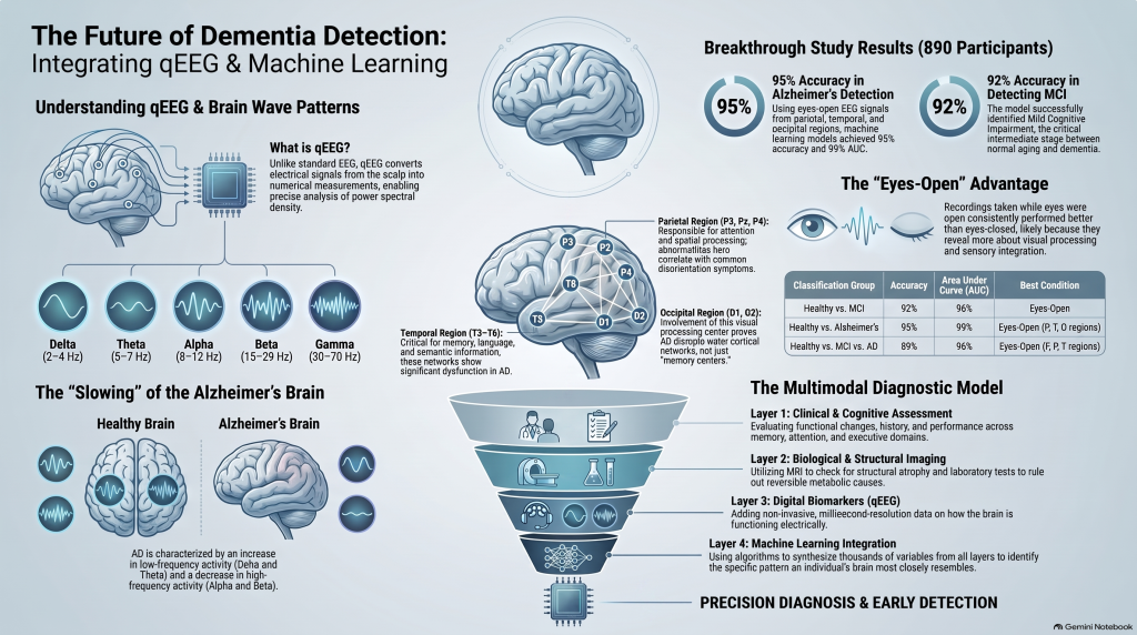
A 2024 study published in Neuropsychiatric Disease and Treatment examined resting-state quantitative EEG (qEEG) from 890 individuals and used machine learning to distinguish healthy controls, mild cognitive impairment (MCI) and Alzheimer’s disease (AD).
The findings are important because they suggest that dementia may produce measurable electrophysiological patterns—and that some brain regions carry more diagnostic information than others.
What Is qEEG?
A conventional EEG records electrical activity generated by the brain through electrodes placed on the scalp.
Quantitative EEG, or qEEG, goes a step further. Instead of relying only on visual inspection of the EEG tracing, the electrical signal is converted into numerical measurements.
One commonly analysed measure is power spectral density, which estimates how much electrical activity is present within different frequency ranges.
In this study, the researchers analysed:
- Delta: 2–4 Hz
- Theta: 5–7 Hz
- Alpha: 8–12 Hz
- Beta: 15–29 Hz
- Gamma: 30–70 Hz
The investigators analysed relative power across 19 EEG channels and grouped these electrodes into frontal, central, temporal, parietal and occipital regions.
What Happens to Brain Waves in Alzheimer’s Disease?
One of the most consistent qEEG findings in Alzheimer’s disease is a general slowing of brain electrical activity.
Compared with healthy individuals, Alzheimer’s disease is commonly associated with:
↑ Delta activity
↑ Theta activity
↓ Alpha activity
and often reduced faster-frequency activity such as beta.
The study reported the same broad pattern, with Alzheimer’s disease showing increased low-frequency activity and reduced alpha activity.
Importantly, this does not mean that increased theta or delta activity automatically indicates Alzheimer’s disease. Similar changes can occur in several neurological, psychiatric and medical conditions.
The value of qEEG may instead come from examining the overall spatial and frequency pattern of brain activity.
The Study: 890 Participants
The researchers analysed:
269 healthy controls
356 people with mild cognitive impairment
265 people with Alzheimer’s disease
Resting EEG was recorded under both:
Eyes-open conditions
and
Eyes-closed conditions
The researchers then converted the EEG signals into quantitative spectral information and trained a Random Forest machine-learning classifier to identify patterns distinguishing the diagnostic groups.
This created four main classification problems:
- Healthy controls vs MCI
- Healthy controls vs Alzheimer’s disease
- Healthy controls vs MCI + Alzheimer’s disease
- Healthy controls vs MCI vs Alzheimer’s disease
Mild Cognitive Impairment: Can qEEG Detect the Intermediate Stage?
Mild cognitive impairment is particularly important because it occupies the clinical space between normal ageing and established dementia.
In the study, the best eyes-open model distinguishing healthy individuals from MCI achieved:
92% accuracy
99% sensitivity
83% specificity
98% negative predictive value
96% AUC
The corresponding eyes-closed model achieved an accuracy of 83%.
This suggests that measurable electrophysiological changes may already be present before full dementia becomes clinically obvious.
Alzheimer’s Disease: The Strongest Classification Result
The strongest performance occurred when distinguishing healthy individuals from patients with Alzheimer’s disease.
Using eyes-open EEG signals from the parietal, temporal and occipital regions, the machine-learning model achieved:
95% accuracy
96% sensitivity
94% specificity
94% positive predictive value
96% negative predictive value
99% AUC
The best eyes-closed model achieved 89% accuracy.
These are impressive research results.
However, they should not be interpreted as meaning that an individual clinical qEEG can diagnose Alzheimer’s disease with 95–99% certainty.
These numbers describe the performance of a machine-learning classifier within this particular research dataset.
Independent validation in broader and more clinically complex populations remains essential.
Eyes Open May Contain More Information Than Expected
One particularly interesting finding was that eyes-open resting EEG consistently performed better than eyes-closed EEG in the machine-learning models.
Traditionally, much resting-state qEEG research has focused on eyes-closed recordings because they are relatively clean and provide strong posterior alpha activity.
But eyes-open EEG may reveal more information related to:
- visual processing
- attention
- sensory integration
- cortical activation
- network responsiveness
The authors suggest that this increased cognitive and sensory processing may help machine-learning models detect abnormalities that are less obvious during eyes-closed recordings.
This is potentially useful because many EEG protocols already collect both eyes-open and eyes-closed data.
Which Brain Regions Were Most Important?
The study identified several regions as particularly informative.
Temporal Region
Electrodes:
T3, T4, T5, T6
The temporal lobes play important roles in:
- memory
- language
- auditory processing
- semantic information
Memory-network dysfunction is central to Alzheimer’s disease, making temporal abnormalities particularly relevant.
Parietal Region
Electrodes:
P3, Pz, P4
Parietal networks contribute to:
- attention
- spatial processing
- navigation
- sensory integration
Visuospatial dysfunction and disorientation are common as dementia progresses.
Occipital Region
Electrodes:
O1, O2
The occipital cortex is primarily involved in visual processing.
The contribution of occipital electrodes reinforces the idea that Alzheimer’s disease is not simply a disorder of a single “memory centre.” It progressively disrupts wider cortical networks.
Central Region
Electrodes:
C3, Cz, C4
These electrodes predominantly reflect activity from sensorimotor regions.
Central channels also provided useful discriminatory information, particularly during eyes-closed recordings.
Dementia Is a Network Disorder
Perhaps the most important conceptual insight from this study is that dementia does not produce a single abnormal EEG electrode.
Instead, disease appears to alter the relationship between frequencies and regions across the brain.
The topographic analyses showed Alzheimer’s disease patients demonstrating:
increased delta and theta power
together with
reduced alpha activity
particularly across temporal and parietal regions.
This fits with the modern understanding of Alzheimer’s disease as a disorder affecting distributed neural networks.
Why Machine Learning Matters
A qEEG recording may generate hundreds or thousands of potential variables.
For example:
frequency × electrode × hemisphere × recording condition × connectivity
A human observer may recognise broad abnormalities, but subtle multidimensional relationships are difficult to process visually.
Machine-learning algorithms can analyse these variables simultaneously and identify combinations that best separate different clinical populations.
The future of EEG biomarkers may therefore move from simply asking:
“Is there excess theta?”
toward asking:
“What overall electrophysiological pattern does this individual’s brain most closely resemble?”
That is a far more sophisticated question.
qEEG Could Become a Digital Biomarker
The emerging field of computational neuroscience is increasingly interested in digital biomarkers—objective physiological measurements that can complement clinical diagnosis.
qEEG has several advantages.
It is:
- non-invasive
- repeatable
- relatively inexpensive
- portable
- capable of measuring brain activity with millisecond temporal resolution
These characteristics make EEG particularly attractive for longitudinal monitoring.
A structural MRI may tell us what the brain looks like.
qEEG potentially adds another dimension:
How is the brain functioning electrically?
But qEEG Does Not Replace a Dementia Evaluation
This distinction is critical.
qEEG remains an adjunctive investigation, not a stand-alone diagnostic test for Alzheimer’s disease.
The authors themselves highlight important limitations of EEG, including:
- relatively poor spatial resolution compared with MRI
- susceptibility to eye movement and muscle artifacts
- environmental electrical interference
- dependence on preprocessing quality
- potential limitations in generalising machine-learning models across populations
They suggest that future approaches may combine EEG with other neuroimaging and multimodal information.
Real-world cognitive assessment must also consider several alternative causes of cognitive symptoms, including:
- depression
- anxiety
- sleep disorders
- medication effects
- vitamin deficiencies
- thyroid disorders
- delirium
- vascular disease
- Parkinsonian disorders
- substance-related conditions
- other neurological illnesses
Clinical context therefore remains indispensable.
The Future Is Multimodal
The most promising approach is unlikely to involve replacing traditional dementia assessment with a single new test.
Instead, future cognitive evaluation may combine several layers of information.
Clinical assessment
What has changed?
How quickly?
How much does it interfere with everyday functioning?
Cognitive testing
Which domains are affected?
Memory?
Attention?
Executive function?
Language?
Visuospatial ability?
MRI or other imaging
Is there structural atrophy, vascular disease or another neurological explanation?
Laboratory and biological markers
Are there reversible metabolic causes or biomarkers suggesting neurodegenerative disease?
qEEG
Are there measurable abnormalities in cortical oscillations or network function?
Machine learning
Can patterns across these different measurements improve diagnostic precision?
This multimodal model is likely to be more clinically useful than relying on any single biomarker.
Key Numbers From the Study
The study included 890 participants and 19 EEG channels.
For healthy controls vs MCI:
92% accuracy using eyes-open EEG.
For healthy controls vs Alzheimer’s disease:
95% accuracy and 99% AUC using eyes-open parietal, temporal and occipital regions.
For healthy controls vs MCI vs Alzheimer’s disease:
89% accuracy and 96% AUC using eyes-open frontal, parietal and temporal regions.
Across the models, eyes-open qEEG generally produced the strongest classification performance.
What Does This Mean for Patients?
For someone experiencing memory problems, qEEG should not be viewed as a shortcut to diagnosis.
The more appropriate question is:
Can qEEG add another objective piece of information to a comprehensive cognitive assessment?
Increasingly, the answer appears to be potentially yes.
The science is moving toward combining clinical symptoms, neuropsychological performance, imaging, biological markers and electrophysiology rather than relying on any single measure.
Conclusion
Quantitative EEG is gradually evolving from simple “brain-wave analysis” into a potential platform for objective neurophysiological measurement and computational classification.
This large study demonstrates that patterns of resting brain electrical activity differ meaningfully between healthy individuals, people with mild cognitive impairment and patients with Alzheimer’s disease.
The strongest signals were not confined to a single location. Temporal, parietal, occipital and central regions all contributed useful information, supporting the idea that cognitive decline reflects disruption of distributed brain networks.
The most interesting future may therefore lie not in asking whether qEEG can independently diagnose dementia, but in determining how much additional information it can provide when combined with clinical examination, cognitive testing, imaging and modern computational analysis.
Comprehensive Cognitive and Dementia Assessment in Chennai
Memory complaints deserve more than a brief screening score.
I provide structured evaluation of memory problems, mild cognitive impairment, dementia and behavioural or psychiatric symptoms associated with cognitive decline, integrating clinical assessment with objective cognitive and neurophysiological measures where clinically appropriate.
Dr. Srinivas Rajkumar T
Senior Consultant Psychiatrist
MD — AIIMS New Delhi
Areas of assessment include:
Memory and cognitive assessment
Mild Cognitive Impairment
Dementia and Alzheimer’s disease
Behavioural and Psychological Symptoms of Dementia (BPSD)
Neuropsychological assessment
Computerised cognitive testing
qEEG-based brain-function assessment where appropriate
Consultations
Apollo Clinic (Opp. Phoenix Market City)
Velachery, Chennai
Appointments: +91 85951 55808
Email: srinivasaiims@gmail.com
Clinical diagnosis remains primary. qEEG and computerized measures are used as adjunctive objective tools when appropriate and should be interpreted alongside history, examination, cognitive assessment, laboratory investigations and neuroimaging.