EEG Microstates in ADHD: Can Brain Network Dynamics Help Predict Treatment Response?
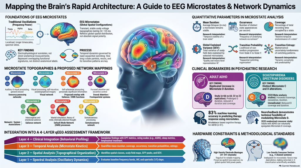 Beyond theta–beta ratios: How quantitative EEG, brain microstates, cognitive testing and artificial intelligence could contribute to the future of precision psychiatry.
Introduction: ADHD Is More Than a Problem of Attention
Imagine two adults diagnosed with Attention-Deficit/Hyperactivity Disorder (ADHD).
Both struggle with procrastination, forgetfulness, distractibility and difficulty completing tasks. Both score similarly on standardized ADHD questionnaires.
Yet their experiences with treatment may be remarkably different.
One experiences substantial improvement in attention and productivity with methylphenidate. Another notices only modest benefits, struggles with side effects or requires a different treatment approach.
Why do people with apparently similar symptoms respond differently?
Part of the answer may lie in the biological and cognitive heterogeneity of ADHD.
ADHD is not a disorder of one isolated brain region or a single neurotransmitter system. It involves complex interactions among networks responsible for attention, executive control, motivation, salience processing and internally directed thought.
Traditionally, quantitative electroencephalography (qEEG) research has examined differences in brain-wave frequencies such as theta, alpha and beta.
However, an emerging field called EEG microstate analysis offers another perspective.
Rather than examining only the frequency of electrical activity, microstate analysis investigates how the spatial organization of brain activity changes from one moment to the next.
This raises an important possibility:
Could the temporal organization of brain activity provide information about ADHD treatment response that conventional qEEG and cognitive testing cannot capture?
It is an exciting question, although the science is still evolving.
1. Understanding Traditional qEEG in ADHD
Electroencephalography records electrical activity generated by populations of neurons. Quantitative EEG applies mathematical and computational methods to these recordings.
Some commonly examined features include:
- Theta activity (4–8 Hz): Often associated with drowsiness, memory processes and aspects of cognitive control.
- Alpha activity (8–13 Hz): Associated with cortical excitability, sensory gating and attentional regulation.
- Beta activity (approximately 13–30 Hz): Involved in motor and cognitive processes, among other functions.
- Individual alpha peak frequency: A measure associated with several aspects of neural function and cognitive performance.
- Functional connectivity: Statistical relationships between electrical signals recorded across brain regions.
Historically, ADHD research paid considerable attention to the theta–beta ratio (TBR).
Some early studies reported elevated theta activity and relatively reduced beta activity in children with ADHD.
This led to considerable optimism that a simple EEG measurement might help identify ADHD objectively.
Unfortunately, subsequent research demonstrated substantial heterogeneity.
Not every individual with ADHD has an elevated theta–beta ratio. EEG findings can vary with age, vigilance, medication exposure, sleep and methodological factors.
Consequently, an elevated theta–beta ratio is not a reliable stand-alone diagnostic marker of ADHD.
The American Academy of Neurology’s practice advisory, reaffirmed in October 2025, continues to caution against using the theta–beta ratio to confirm ADHD outside appropriately designed research.
This does not mean qEEG research lacks value.
Instead, it suggests that a complex neurodevelopmental disorder cannot be adequately explained by one spectral ratio.
We may need to study not only the frequencies of brain activity but also how electrical activity is spatially and temporally organized.
2. What Are EEG Microstates?
Even during quiet wakefulness, the brain undergoes continuous changes in electrical activity.
Interestingly, these changes are not completely random.
For brief periods, the overall spatial distribution of electrical potentials across the scalp remains approximately stable before transitioning to another configuration.
These relatively stable configurations are called EEG microstates.
A typical microstate lasts approximately 50–120 milliseconds.
This means that the brain can transition between several different electrical configurations within a single second.
Researchers have identified recurring patterns that appear across many individuals.
Four classical configurations are conventionally labeled:
- Microstate A
- Microstate B
- Microstate C
- Microstate D
More recent studies also examine additional classes.
Microstates are identified through computational analysis of multichannel EEG recordings. The analysis typically involves extracting scalp topographies, clustering recurring patterns and measuring their temporal characteristics.
Several parameters can be quantified.
| Microstate parameter | What it measures |
|---|---|
| Duration | How long a particular configuration persists |
| Occurrence | How frequently that configuration appears |
| Coverage | What proportion of the recording it occupies |
| Transition probability | How often one microstate changes into another |
| Global explained variance | How well the identified maps represent the recorded EEG |
Microstates have sometimes been described metaphorically as the atoms of thought.
Although memorable, this expression should not be interpreted literally.
A microstate cannot identify what someone is thinking. It reflects a recurring spatial configuration of electrical activity rather than a specific thought, emotion or memory.
3. What Do Microstates Tell Us About Brain Networks?
One of the most important developments in neuroscience has been the recognition that cognitive functions emerge from interactions among distributed brain networks.
Three networks are particularly interesting in ADHD.
The Default Mode Network: Internally Directed Thought
The Default Mode Network (DMN) is associated with spontaneous thinking, autobiographical memory, self-referential processing and mental simulation.
It involves regions including the medial prefrontal cortex, posterior cingulate cortex, precuneus and angular gyrus.
During demanding externally oriented tasks, activity in parts of this network often decreases relative to rest.
However, the DMN is not simply a distraction network. It performs essential functions in memory, imagination and internally directed cognition.
In ADHD, researchers have investigated whether altered interactions between internally directed processing and task-positive networks contribute to attentional inconsistency.
The Salience Network: Detecting What Matters
The Salience Network involves regions such as the anterior insula and dorsal anterior cingulate cortex.
It contributes to detecting behaviorally relevant information and coordinating activity across other networks.
For example, when a person is concentrating on a task and an unexpected event demands attention, salience-related processes help determine whether the event should interrupt ongoing activity.
Attention and Executive Control Networks
The Frontoparietal Control Network and Dorsal Attention Network contribute to working memory, executive functioning and goal-directed attention.
Their coordination is particularly important during sustained cognitive tasks.
ADHD may involve disturbances in how these distributed systems interact across time.
Where do microstates fit?
Research combining EEG, source localization and functional MRI has identified associations between certain scalp microstate configurations and distributed functional networks.
Microstate D, for example, has frequently been associated with attention-related and executive processes.
Microstate C has been linked to self-referential and personally significant information processing, with some studies proposing salience-related functions.
However, these relationships are not definitive.
A microstate cannot be equated directly with a specific functional network.
In particular, microstate C is not universally synonymous with the salience network, and microstate D is not a direct measurement of executive control.
The functional review by Tarailis and colleagues (2024) emphasized these important complexities.
The value of microstate analysis lies in measuring rapidly changing electrical configurations that may relate to the activity of distributed neuronal systems.
4. What Does Microstate Research Actually Show in ADHD?
The scientific evidence is increasingly interesting.
Study 1: Microstate D Duration in Adult ADHD
Férat and colleagues, in a study published in Biological Psychiatry: Cognitive Neuroscience and Neuroimaging in 2022, investigated resting-state microstates across two independent adult datasets.
The first included 66 adults with ADHD and 66 healthy controls.
The second included 22 adults with ADHD and 22 controls.
The researchers found that the canonical spatial microstate patterns were broadly preserved in ADHD.
However, their temporal characteristics differed.
Most notably, microstate D exhibited longer mean duration in adults with ADHD, a finding reproduced across both datasets.
Microstate A also showed reduced duration and coverage in the larger dataset.
Some microstate characteristics were associated with sleep disturbance.
What does this mean?
One interpretation is that ADHD may involve changes in how persistently certain electrical configurations dominate brain activity.
It is possible that some configurations become relatively more stable or less flexible.
However, the finding does not establish whether longer microstate D duration represents a primary abnormality, a compensatory mechanism or a consequence of another physiological process.
Nevertheless, the replicated finding suggests that temporal EEG organization deserves attention.
Study 2: Microstate Differences in Children With ADHD
Luo and colleagues examined high-density EEG in 161 participants, including children with predominantly inattentive ADHD, combined-presentation ADHD and healthy controls.
The study reported:
- Reduced microstate C coverage in ADHD.
- Increased duration and contribution of microstate D.
- Additional microstate differences between ADHD presentations.
These findings broadly support the possibility that ADHD involves altered temporal organization of large-scale electrical activity.
However, microstate measurements are not sufficiently validated to differentiate ADHD presentations in routine clinical practice.
Study 3: The 2025 Systematic Review
Berchio and colleagues reviewed 13 studies investigating EEG microstates in ADHD.
The review described recurring findings involving the temporal dynamics of microstates C and D.
It also identified evidence from task-based studies suggesting that microstate organization may differ during processes involving attention and inhibitory control.
However, the literature remained heterogeneous.
Differences in recording systems, participant characteristics, medication exposure, microstate clustering methods and analytical approaches limit the generalizability of these findings.
This is particularly important when interpreting individual patient recordings.
5. A Major Question: Does Microstate Analysis Add Anything Beyond CPT?
The Continuous Performance Test (CPT) is commonly used in ADHD assessment and research.
It measures aspects of attention and response control, including omission errors, commission errors, reaction-time variability and sustained performance.
CPT findings are not specific enough to diagnose ADHD independently, but they provide a complementary behavioral measurement.
This creates an important scientific question:
If we already have clinical assessments and objective cognitive testing, does adding EEG microstate analysis improve our ability to understand ADHD?
A study published by Wei and colleagues in BMC Psychiatry in September 2026 directly addressed part of this question.
The researchers examined 121 children aged 6–15 years, including 63 with ADHD and 58 typically developing controls.
Using resting-state EEG, they found reduced occurrence of microstate C and reduced C-to-D transition probability in the ADHD group.
The study then evaluated machine-learning classification models.
The results were interesting:
| Classification model | Area under ROC curve (AUC) |
|---|---|
| EEG microstates alone | 0.744 |
| IVA-CPT alone | 0.913 |
| Microstates + IVA-CPT | 0.930 |
At first glance, the combined approach appeared promising.
However, adding microstate information did not produce a statistically significant improvement over CPT alone.
These results came from internal cross-validation in one dataset, not independent clinical validation.
The study offers an important lesson.
Adding more biological measurements does not automatically produce a better clinical assessment.
Every proposed new biomarker should demonstrate that it contributes useful information beyond existing methods.
This principle is central to the development of reliable precision psychiatry.
6. Why Treatment Prediction May Be More Valuable Than Diagnosis
Much of the early enthusiasm surrounding qEEG focused on developing objective tests capable of identifying psychiatric diagnoses.
However, ADHD is clinically heterogeneous.
Two people may meet the same diagnostic criteria while differing considerably in executive functioning, emotional regulation, sleep patterns, cognitive performance and medication response.
This creates an opportunity to ask a more clinically meaningful question.
Instead of asking only:
Can EEG identify ADHD?
We could ask:
Can EEG help predict which treatment is most likely to benefit a particular person with ADHD?
This would represent a shift from diagnostic classification toward treatment stratification.
Consider methylphenidate, a commonly prescribed stimulant medication for ADHD.
Its clinical effects vary across individuals.
Some experience improvements in sustained attention, task initiation and inhibitory control.
Others may respond incompletely, experience adverse effects or require treatment adjustments.
There is currently no validated EEG microstate profile that can reliably predict an individual’s response to methylphenidate.
Nevertheless, microstate research creates several testable hypotheses.
For example:
- Does baseline microstate D duration correlate with subsequent improvement in executive functioning?
- Are particular microstate transition patterns associated with better CPT outcomes following treatment?
- Do successful treatments change microstate organization over time?
- Are EEG changes associated with meaningful functional improvement rather than simply changes in test scores?
These questions require prospective studies with careful control of medication timing, sleep, vigilance and other confounding factors.
Most importantly, improvements in symptoms and functioning must remain the primary outcomes.
7. What About Methylphenidate and EEG Changes?
There is already a substantial literature examining how methylphenidate affects EEG.
A systematic review published in September 2026 by Quinn and colleagues examined 44 studies involving acute methylphenidate administration in ADHD.
The most consistent finding concerned increased P3 amplitude, an event-related potential associated with aspects of cognitive processing.
However, the authors concluded that existing EEG evidence does not establish a validated treatment-response biomarker for methylphenidate.
Microstate-specific treatment evidence remains much more limited.
An additional randomized crossover investigation published in December 2025 examined microstate D neurofeedback in 19 adults with ADHD.
Participants demonstrated the ability to increase microstate D coverage during neurofeedback targeting upregulation.
However, downregulation was not significant, and the study did not demonstrate short-term clinical improvement.
This suggests that particular EEG microstate characteristics may be experimentally modifiable.
It does not demonstrate that manipulating a microstate improves ADHD symptoms.
These findings reinforce the importance of distinguishing neurophysiological change from clinical benefit.
8. Could qEEG and Cognitive Testing Identify Treatment-Relevant ADHD Subgroups?
A particularly relevant development appeared in the Journal of the American Academy of Child & Adolescent Psychiatry in 2026.
Leikauf and colleagues investigated whether ADHD subgroups defined by cognitive performance could have distinct physiological profiles and medication responses.
The researchers identified and replicated two cognitive subgroups in separate clinical samples.
One group was characterized primarily by difficulties in inhibitory control, while the other showed a different profile centered on sustained attention.
The inhibitory-control subgroup exhibited differences in resting qEEG spectral activity, resting heart rate and anxiety symptoms.
Importantly, this subgroup demonstrated a greater response to atomoxetine than the comparison subgroup.
The findings offer preliminary support for the possibility that cognitive and physiological measurements may help identify treatment-relevant ADHD heterogeneity.
An important distinction: this study examined conventional qEEG spectral features, not resting-state EEG microstates as a treatment predictor.
Nevertheless, it provides a useful conceptual direction.
Future studies could investigate whether microstate dynamics add predictive information beyond the cognitive and spectral variables already examined.
That would be a meaningful next step rather than simply adding another complex EEG feature to an existing model.
9. Integrating qEEG, Microstates, CPT and Artificial Intelligence
I believe the greatest opportunity for objective psychiatric assessment lies in intelligently combining complementary sources of information.
No single measurement adequately captures the complexity of ADHD.
A comprehensive research framework could involve four levels.
Level 1: Clinical Phenotyping
A structured psychiatric assessment establishes the clinical context.
This includes developmental history, functional impairment, ADHD symptom patterns, sleep, anxiety, mood symptoms, substance use and relevant medical conditions.
Standardized ADHD scales can provide structured symptom measurements.
Clinical assessment also helps distinguish ADHD from secondary causes of inattention.
Level 2: Cognitive and Behavioral Testing
Computerized attention testing can measure sustained attention, reaction-time variability and response inhibition.
Additional neuropsychological measures may be included where appropriate.
These tests provide objective behavioral information, although their findings are influenced by motivation, fatigue, sleep and testing conditions.
Level 3: Neurophysiological Assessment
Appropriate multichannel qEEG recordings may provide several complementary measurements.
Conventional analysis can examine spectral power, individual alpha frequency and other validated signal features.
Research-grade microstate analysis can additionally examine:
- Mean microstate duration
- Frequency of occurrence
- Time coverage
- Global explained variance
- Transition probabilities
- Sequence organization
Connectivity analysis could form another research dimension when the recording montage and methodology are suitable.
These physiological features describe aspects of brain activity, not psychiatric diagnoses themselves.
Level 4: Longitudinal Treatment Outcomes
The most meaningful step is to connect baseline assessments with what happens after treatment.
Follow-up may examine improvement in ADHD symptoms, daily functioning, cognitive performance and tolerability.
For example, a research program could investigate changes following methylphenidate or atomoxetine while accounting for medication dose and timing.
A predictive model would then attempt to determine whether baseline clinical, cognitive and neurophysiological characteristics provide useful information about subsequent outcomes.
Rather than assuming that adding EEG improves prediction, researchers should compare models systematically.
A clinical-only model should be compared with clinical plus CPT, clinical plus qEEG, and finally a model incorporating microstate features.
The additional measurements would be considered useful only if they improved prediction in properly validated data and offered meaningful clinical benefits.
10. Why Artificial Intelligence Matters—And Why Caution Is Essential
EEG signals contain substantial amounts of multidimensional information.
Machine-learning methods can identify statistical relationships among variables that may be difficult to detect using conventional analysis.
However, machine learning also introduces important risks.
Small research datasets can produce apparently impressive predictive accuracy that disappears when a model is tested on independent patients.
A model may inadvertently learn differences in recording conditions, age, medication exposure or technical artifacts rather than meaningful characteristics of ADHD.
Repeated EEG recordings from the same participant can also create data leakage if observations from that participant appear in both training and test datasets.
Reliable development therefore requires careful preprocessing, participant-level data separation, appropriate cross-validation, calibration and external validation.
Ideally, predictive models should also demonstrate that they improve clinical decisions rather than merely increasing statistical accuracy.
Artificial intelligence should be used to test biological hypotheses and support clinical reasoning—not to transform uncertain measurements into unjustified diagnostic certainty.
11. Can Low-Cost EEG Devices Contribute to This Research?
One of the most significant barriers to widespread neurophysiological research is the cost of equipment, infrastructure and repeated measurements.
Portable EEG devices have made it increasingly feasible to record electrical activity outside traditional laboratories.
This creates opportunities for longitudinal investigations of attention, sleep, cognitive performance and treatment-related physiological changes.
However, the distinction between spectral qEEG and microstate mapping is important.
Classical microstate analysis requires sufficient spatial coverage of the scalp to identify recurring electrical configurations.
Four-channel wearable systems, including devices with electrodes concentrated over frontal and temporal areas, have substantial limitations for reconstructing conventional whole-scalp microstates.
A more comprehensive electrode montage is preferable for reliable research-grade microstate analysis.
For low-cost longitudinal EEG research, it may therefore be more appropriate to begin with spectral measures that have been validated for the specific device, while conducting microstate investigations using separate multichannel recordings.
The aim should be to balance accessibility with scientific validity.
An affordable measurement becomes clinically valuable only when it is sufficiently reliable, interpretable and useful.
12. From Brain Mapping to Precision Psychiatry
The future of ADHD assessment may not lie in finding a single EEG signature shared by everyone with the disorder.
It may instead lie in understanding why individuals with similar clinical symptoms have different underlying physiological and cognitive characteristics.
This could eventually allow clinicians to ask increasingly specific questions.
Which patients are most likely to benefit from a particular medication?
Which cognitive difficulties remain despite improvement in core ADHD symptoms?
Can physiological measurements help identify early treatment-associated changes?
Can repeated recordings distinguish meaningful longitudinal patterns from temporary fluctuations in alertness or sleep?
And most importantly, can additional physiological information improve outcomes enough to justify the cost and complexity of testing?
These are the questions that should guide the next generation of psychiatric research.
Microstate analysis offers a valuable experimental window into the rapidly changing spatial organization of brain electrical activity.
But it is only one component of a much broader scientific challenge.
Conclusion: The Future of ADHD Assessment Is Multidimensional
ADHD cannot be fully understood through symptoms alone, but neither can it be reduced to a single EEG measurement.
Traditional qEEG describes important aspects of brain oscillations.
Microstate analysis adds information about recurring electrical configurations and their temporal dynamics.
Cognitive testing examines how attention and executive control are expressed through behavior.
Clinical assessment places these findings within the individual’s developmental, psychological and functional context.
Artificial intelligence may eventually help integrate these different dimensions.
The central challenge is to demonstrate that this integration leads to more accurate predictions, better treatment decisions and meaningful improvements in patients’ lives.
The future of ADHD research may depend less on discovering one perfect diagnostic biomarker and more on understanding which combination of clinical, cognitive and neurophysiological measurements can guide genuinely personalized treatment.
That, to me, is the more important direction for the future of precision psychiatry.
About the Author: Advanced ADHD Assessment in Chennai
Dr. Srinivas Rajkumar T is a Senior Consultant Psychiatrist in Chennai, with an MD in Psychiatry from the All India Institute of Medical Sciences (AIIMS), New Delhi, DNB Psychiatry and an MBA from BITS Pilani.
His clinical and academic interests include adult ADHD, quantitative EEG, cognitive neuroscience, neurophysiology, neuromodulation and the application of emerging technologies to evidence-based psychiatric care.
He has a particular interest in integrating structured psychiatric assessments, computerized attention testing and neurophysiological measurements to understand individual differences in ADHD.
Attention Clinic — Attention. Understood.
At the Attention Clinic, Chennai, ADHD assessment is approached through a comprehensive, specialist-led model.
Depending on individual clinical requirements, assessment may include:
- Detailed psychiatric and developmental evaluation
- Standardized ADHD rating scales
- Computerized Continuous Performance Testing (CPT)
- Cognitive and executive-function assessment
- qEEG-assisted evaluation where appropriate
- Individualized medication planning
- ADHD-focused psychological therapy
- Executive-function strategies and long-term follow-up
The clinic emphasizes combining careful clinical reasoning with objective measurements wherever these genuinely add value.
Our goal is not simply to identify ADHD, but to understand the difficulties affecting each individual and develop a practical, personalized treatment plan.
Research techniques such as EEG microstate analysis remain investigational and are not presented as established clinical diagnostic services.
Dr. Srinivas Rajkumar T
Senior Consultant Psychiatrist
MD Psychiatry (AIIMS, New Delhi) | DNB Psychiatry
Attention Clinic — Comprehensive 360° ADHD Care
📍 Apollo Clinic, Velachery, Opposite Phoenix Marketcity, Chennai
📞 Appointments: +91 85951 55808
🌐 Website: https://www.srinivasaiims.com
🔗 Learn more about Attention Clinic – Comprehensive ADHD Care in Chennai