Neuropsychological Assessment by Cognitive Domain: Understanding What Each Test Reveals About the Brain
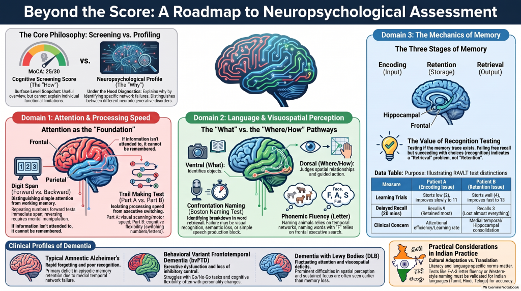 Introduction: Beyond the Cognitive Screening Score
A 68-year-old patient presents with progressive forgetfulness. The Montreal Cognitive Assessment (MoCA) score is 25/30. Another patient scores 19/30 but remains relatively independent in everyday activities. A third patient scores 27/30 despite experiencing significant difficulty recognising objects and navigating familiar surroundings.
How should these findings be interpreted?
The answer lies in understanding that cognition is not a single ability. It consists of multiple interacting processes, including attention, processing speed, language, executive functioning, visuospatial perception, and episodic memory.
A global cognitive screening score provides a useful overview of cognitive performance, but it cannot fully explain the nature of an individual’s cognitive difficulties.
Two patients with identical MoCA scores may have entirely different cognitive profiles, underlying disorders, functional limitations, and treatment needs.
This is where detailed neuropsychological assessment becomes particularly valuable.
Neuropsychological tests examine specific cognitive abilities through structured tasks. By studying patterns of performance across multiple tests, clinicians can identify the cognitive processes most affected, distinguish primary impairments from secondary difficulties, and develop a more meaningful clinical formulation.
This approach is particularly important in dementia assessment because not all neurodegenerative disorders begin with memory impairment. Some patients initially experience language difficulties, behavioural changes, executive dysfunction, or visuospatial problems.
The central principle is simple:
A cognitive screening score tells us how a patient performed. A neuropsychological profile helps us understand why.
This article explores important neuropsychological tests organised by cognitive domain, explains the brain networks involved, and discusses how the findings can contribute to dementia diagnosis and clinical management.
1. Attention: The Foundation of Cognitive Functioning
Attention is the ability to select, maintain, and manipulate information while filtering out irrelevant stimuli.
It is fundamental to nearly every other cognitive process.
For example, a person cannot remember a conversation that was never adequately attended to in the first place.
Similarly, difficulty maintaining attention may interfere with language comprehension, planning, learning, and everyday decision-making.
Attention is not a single function. It includes immediate attention span, sustained attention, selective attention, divided attention, working memory, and inhibitory control.
Different neuropsychological tests examine these components.
1.1 Immediate Attention Span: Digit Span
The Digit Span test is commonly included in the Wechsler Adult Intelligence Scale and other neuropsychological batteries.
The examiner reads a sequence of numbers aloud, and the patient repeats them according to specific instructions.
There are three commonly used versions.
Digit Span Forward requires the patient to repeat numbers in the same order.
For example, the examiner says: 4, 8, 2, 9.
The patient responds: 4, 8, 2, 9.
This primarily examines immediate auditory attention span and the capacity to maintain verbal information briefly.
Digit Span Backward requires the patient to repeat the numbers in reverse order.
If the examiner says 4, 8, 2, 9, the patient should respond 9, 2, 8, 4.
This requires not only maintaining information but also mentally manipulating it.
Consequently, Digit Span Backward places greater demands on working memory and executive control.
Digit Span Sequencing requires the patient to rearrange the digits into ascending numerical order.
This additionally engages sequencing and mental organisation.
Brain regions involved:
Digit Span performance depends on distributed frontoparietal working-memory networks.
Dorsolateral prefrontal regions contribute to executive manipulation and control of information.
Posterior parietal regions contribute to maintaining information in working memory.
Verbal processing networks also contribute to auditory digit span.
Clinical significance:
A patient who performs relatively well on Digit Span Forward but poorly on Digit Span Backward may have difficulty manipulating information despite relatively preserved immediate attention span.
Such a pattern can occur in several neurological and psychiatric conditions.
However, poor Digit Span Backward performance does not independently establish frontal lobe dysfunction.
Hearing impairment, anxiety, fatigue, language difficulties, and educational background may also influence performance.
In dementia assessment, Digit Span can help determine whether an apparent memory complaint is partly related to impaired attention or working memory.
1.2 Sustained Attention: MoCA Letter-A Vigilance Task
Sustained attention refers to the ability to maintain focus over time.
The Letter-A Vigilance Task in the MoCA provides a brief assessment of this ability.
The examiner reads a sequence of letters, and the patient is instructed to tap whenever the letter A is heard.
The patient must continuously monitor the sequence, identify the target, and avoid responding to other letters.
Errors may include missed targets or responses to incorrect letters.
Missed targets may suggest attentional lapses.
Responses to non-target letters may indicate difficulties with response control, although misunderstanding or hearing problems must also be considered.
The MoCA vigilance task is a brief screening item. Its limited length means it cannot characterise sustained attention as comprehensively as a dedicated attention battery.
Brain networks involved:
Sustained attention depends on frontal and parietal networks, including right-lateralised attention systems, together with thalamic and arousal-related systems.
Clinical significance:
Attention assessment is particularly important when evaluating patients with fluctuating cognition.
Fluctuating attention may occur in delirium and dementia with Lewy bodies.
However, a single abnormal vigilance task cannot establish either diagnosis.
The clinical history, pattern of fluctuation, neurological findings, and possible medical contributors remain essential.
1.3 Continuous Performance Tests
Continuous Performance Tests, commonly called CPTs, provide a more detailed assessment of sustained attention and response control.
During a typical CPT, the patient watches or listens to a continuous series of stimuli.
The patient must respond to designated targets and withhold responses to non-targets.
Depending on the specific CPT, the assessment may generate several measures.
Omission errors occur when the patient fails to respond to a target.
Commission errors occur when the patient responds to a non-target.
Reaction time measures how quickly the patient responds.
Reaction-time variability measures the consistency of responses over time.
Some tests also examine changes in performance across different task conditions or longer periods of testing.
These measures provide information about different aspects of attentional performance.
For example, a patient may respond slowly but consistently.
Another patient may respond quickly but make frequent commission errors.
A third may demonstrate substantial variability, alternating between periods of adequate performance and attentional lapses.
These patterns may have different clinical implications.
Clinical significance:
CPTs are commonly used in the assessment of attention-related difficulties, including ADHD.
They may also provide useful supplementary information in neurological and neuropsychological assessment.
However, CPT abnormalities are not specific to ADHD, dementia, or any other single disorder.
Sleep deprivation, depression, anxiety, medications, neurological illness, sensory limitations, and inadequate task understanding can influence performance.
CPT results should therefore be interpreted as part of a broader clinical assessment rather than as a standalone diagnostic test.
1.4 Response Inhibition: Motor Go/No-Go Task
Response inhibition refers to the ability to suppress an automatic or inappropriate response.
The Motor Go/No-Go Task examines this ability.
The patient is instructed to respond to one stimulus and withhold the response to another.
For example, the examiner may ask the patient to tap once when a particular signal is presented but remain still when another signal appears.
The task requires the patient to understand the instructions, maintain the rules, monitor incoming stimuli, and suppress inappropriate responses.
A patient with impaired inhibitory control may repeatedly respond when instructed not to.
Brain networks involved:
Response inhibition involves frontostriatal circuits, including the inferior frontal cortex, presupplementary motor area, basal ganglia, and associated control networks.
Clinical significance:
Impaired inhibition may be observed in patients with frontal-system dysfunction, certain movement disorders, acquired brain injuries, and some neurodegenerative conditions.
In behavioural variant frontotemporal dementia, reduced inhibitory control may accompany socially inappropriate behaviour, impulsivity, or difficulty regulating actions.
However, a poor Go/No-Go score alone does not establish a frontal neurodegenerative syndrome.
Attention, comprehension, motor ability, and the broader behavioural history must be evaluated.
1.5 Executive Attention: Trail Making Test Part B
The Trail Making Test is among the most widely used neuropsychological measures of attention, processing speed, and executive functioning.
Part B requires the patient to connect numbers and letters in alternating ascending order.
For example:
1 – A – 2 – B – 3 – C – 4 – D.
The patient must maintain two sequences simultaneously while repeatedly switching between them.
This requires cognitive flexibility, divided attention, sequencing, and monitoring.
The time taken to complete the task and the types of errors made are important.
A patient who repeatedly connects numbers without switching to letters may have difficulty maintaining or implementing the alternating rule.
Another patient may understand the rule but perform slowly because of reduced processing speed.
A third may struggle because of poor visual scanning or limited familiarity with the alphabet.
Brain networks involved:
Trail Making Test Part B depends on distributed frontoparietal and frontostriatal networks, including dorsolateral prefrontal regions.
Clinical significance:
Executive difficulties may be prominent in vascular cognitive impairment, frontal-system disorders, Parkinsonian syndromes, and several psychiatric conditions.
Importantly, Part B is not a pure measure of executive functioning.
It also depends on processing speed, visual scanning, motor functioning, and familiarity with numbers and letters.
Comparing performance with Trail Making Test Part A can help identify some of these contributing factors.
However, even derived measures such as the Part B–Part A difference or B/A ratio do not completely isolate executive function.
2. Processing Speed: How Efficiently the Brain Handles Information
Processing speed refers to the rate at which an individual can perceive information, perform relatively simple cognitive operations, and generate responses.
It is a fundamental component of cognitive efficiency.
A person with reduced processing speed may understand information accurately but require additional time to process it.
This can affect conversations, decision-making, learning, and everyday activities.
Processing speed is particularly relevant in vascular cognitive impairment and other disorders involving distributed brain networks.
2.1 Trail Making Test Part A
Trail Making Test Part A requires the patient to connect numbered circles in ascending order.
For example:
1 – 2 – 3 – 4 – 5.
The task appears straightforward, but successful performance requires visual scanning, attention, sequencing, motor coordination, and processing speed.
The time required to complete the task is typically recorded.
A prolonged completion time may reflect reduced processing speed.
However, visual impairment, motor slowing, poor familiarity with the task, or attentional difficulties may also contribute.
Brain networks involved:
Performance depends on distributed visual, parietal, frontal, and motor systems.
It cannot be attributed to a single brain region.
Clinical significance:
Part A provides useful information when interpreting more complex tasks such as Trail Making Test Part B.
If a patient performs slowly on both tasks, general processing speed or visual-motor difficulties may contribute to the poor Part B performance.
If Part B is disproportionately impaired, additional executive demands may be relevant.
2.2 Coding Tests
Coding tests are included in instruments such as the Wechsler Adult Intelligence Scale and the Repeatable Battery for the Assessment of Neuropsychological Status (RBANS).
In a typical coding task, the patient must rapidly associate numbers with symbols according to a reference key.
The task requires sustained attention, visual scanning, rapid learning of symbol-number associations, and motor output.
Coding tests are sensitive to changes in cognitive efficiency.
However, their sensitivity also means that poor performance is not specific to a particular disease.
Clinical significance:
Slowed coding performance may occur in vascular cognitive impairment, depression, neurological disease, sleep disorders, and medication-related cognitive slowing.
Motor impairment can also substantially affect performance.
For this reason, coding scores should be interpreted alongside other measures and the patient’s clinical circumstances.
A patient with Parkinsonian motor slowing, for example, may perform poorly on a timed written coding task even when some underlying cognitive abilities remain relatively preserved.
The examiner must determine how much of the difficulty is cognitive and how much is motor.
3. Language: Understanding, Producing, and Organising Meaning
Language is a complex cognitive system involving speech production, comprehension, naming, repetition, reading, and writing.
Language difficulties are particularly important in the assessment of primary progressive aphasia, stroke, and atypical Alzheimer’s disease.
Not all language impairments are the same.
A patient may understand a word but be unable to retrieve it.
Another may produce fluent speech but have difficulty understanding the meaning of words.
A third may understand language relatively well but struggle to produce grammatically organised sentences.
These patterns suggest dysfunction in different components of the language network.
3.1 Comprehensive Aphasia Assessment
The Boston Diagnostic Aphasia Examination and Western Aphasia Battery-Revised are structured language assessment batteries.
They examine multiple language abilities rather than focusing on one task.
Important components include spontaneous speech, auditory comprehension, naming, repetition, reading, and writing.
Spontaneous speech is assessed for fluency, speech rate, grammar, articulation, word choice, and meaningful content.
Auditory comprehension examines the ability to understand spoken words, commands, and sentences.
Naming evaluates the ability to retrieve words for objects, actions, or concepts.
Repetition assesses the ability to repeat words, phrases, and sentences.
Reading and writing examine additional components of language processing.
These assessments help identify the nature and severity of language impairment.
Clinical significance:
A comprehensive language profile can help distinguish different forms of aphasia.
For example, a patient with fluent speech but impaired single-word comprehension may have a different syndrome from a patient with effortful, grammatically simplified speech.
The distinction is particularly relevant when evaluating primary progressive aphasia.
However, a language battery identifies the clinical language syndrome. It does not independently establish the underlying molecular pathology.
3.2 Naming: Boston Naming Test
The Boston Naming Test examines confrontation naming.
The patient is shown pictures of objects and asked to name them.
Naming requires several cognitive processes to work together.
First, the patient must visually perceive the object.
Second, the object must be recognised.
Third, semantic knowledge about the object must be accessed.
Fourth, the appropriate word must be retrieved.
Finally, the word must be produced.
Difficulty at any stage may interfere with naming.
Consider a patient who is shown a picture of a camel but cannot name it.
Several explanations are possible.
The patient may not recognise the picture.
The patient may recognise the animal but have difficulty retrieving its name.
The patient may have lost some of the semantic knowledge associated with the animal.
Alternatively, speech production difficulties may prevent the correct response.
The examiner can explore these possibilities by asking the patient to describe the object, identify it from alternatives, or respond to semantic and phonemic cues.
For example, if the patient can describe the animal accurately and produces its name after hearing the first sound, lexical retrieval difficulty may be contributing.
If the patient cannot identify the animal or explain what it is, a broader semantic impairment may need to be considered.
Brain networks involved:
Naming involves distributed language networks, particularly dominant temporal regions, with contributions from visual recognition and frontal retrieval systems.
Clinical significance:
Naming impairment may occur in Alzheimer’s disease, primary progressive aphasia, focal brain lesions, and other neurological conditions.
However, an abnormal Boston Naming Test score is not sufficient to distinguish these disorders.
The nature of the errors and performance on other language tasks are essential.
3.3 Word Comprehension: Peabody Picture Vocabulary Test
The Peabody Picture Vocabulary Test, Fourth Edition, assesses receptive vocabulary.
The examiner says a word, and the patient selects the corresponding picture from several alternatives.
Unlike the Boston Naming Test, the patient is not required to produce the word.
This distinction helps separate word retrieval from word comprehension.
A patient who cannot say the word elephant may still correctly identify the animal when the examiner says its name.
This suggests that some semantic knowledge is preserved despite difficulty producing the word.
In contrast, a patient with impaired semantic knowledge may struggle to identify the correct picture even when the word is provided.
Brain networks involved:
Word comprehension depends on distributed language and semantic networks, including important contributions from the dominant temporal cortex.
Clinical significance:
Receptive vocabulary assessment may help characterise semantic impairment.
It can be particularly informative when evaluating patients with suspected semantic variant primary progressive aphasia.
However, the Peabody Picture Vocabulary Test is not a dementia-specific diagnostic instrument.
Performance is influenced by language exposure, educational background, cultural familiarity, and premorbid vocabulary.
Appropriate language-specific assessment is therefore essential.
3.4 Semantic Fluency: Category List Generation
Semantic fluency tasks require the patient to generate as many words as possible within a category during a specified time.
A commonly used example is animal naming.
The examiner asks the patient to name as many animals as possible in one minute.
Other categories may include vegetables, fruits, or other familiar groups.
Successful performance requires access to semantic knowledge, lexical retrieval, processing speed, and executive search strategies.
The total number of words generated is important, but the organisation of responses can provide additional information.
For example, a patient may begin with farm animals, move to wild animals, and then name birds.
This illustrates clustering and switching.
Clustering refers to producing several related words within a semantic subcategory.
Switching refers to moving between subcategories.
Reduced clustering may suggest difficulty accessing or organising semantic information.
Reduced switching may suggest difficulty changing retrieval strategies.
However, these measures are influenced by several cognitive systems and should not be considered anatomically specific.
Clinical significance:
Semantic fluency may be disproportionately impaired in disorders affecting semantic knowledge, including semantic variant primary progressive aphasia.
It may also be impaired in Alzheimer’s disease and other conditions involving temporal, frontal, or distributed cognitive networks.
The pattern of errors, clustering, switching, and performance on complementary language tests helps clarify the underlying difficulty.
3.5 Phonemic Fluency: F-A-S Test
Phonemic fluency requires the patient to generate words beginning with specified letters.
The F-A-S Test commonly uses the letters F, A, and S in separate timed trials.
The patient must generate words beginning with the designated letter while following rules that typically exclude proper nouns and simple variations of the same word.
Unlike category fluency, phonemic fluency does not primarily rely on retrieving items from a familiar semantic category.
It requires strategic lexical search, cognitive flexibility, response monitoring, and inhibition of inappropriate responses.
Brain networks involved:
Phonemic fluency depends on distributed left-hemisphere language and executive networks, including frontal contributions.
Clinical significance:
Poor phonemic fluency may occur in patients with executive dysfunction or language impairment.
Comparing phonemic and semantic fluency can provide useful information about the relative contribution of executive search and semantic knowledge.
However, the comparison should not be interpreted as a simple frontal-versus-temporal diagnostic test.
Both tasks engage overlapping networks.
An additional challenge arises in multilingual populations.
The F-A-S Test was developed for English-language assessment. Its original norms cannot automatically be applied to Tamil, Hindi, Telugu, or other languages.
Appropriately validated language-specific measures and norms should be used wherever possible.
4. Visuospatial Perception: Recognising and Interpreting the Visual World
Visuospatial cognition enables individuals to recognise objects, judge spatial relationships, navigate environments, interpret visual scenes, and construct meaningful representations.
These abilities are particularly important in everyday activities such as reading, driving, dressing, finding objects, and moving through familiar surroundings.
Visuospatial cognition includes several related but distinct functions.
Object perception involves identifying what an object is.
Spatial perception involves judging where objects are and how they relate to one another.
Spatial representation involves constructing or mentally manipulating spatial information.
Different neuropsychological tests examine these components.
A useful anatomical framework distinguishes two major visual processing pathways.
The ventral visual pathway extends from occipital regions toward temporal cortex and is particularly important for object recognition and visual identity.
It is commonly described as the what pathway.
The dorsal visual pathway extends from occipital regions toward parietal cortex and contributes to spatial processing and visually guided action.
It is often described as the where or how pathway.
These pathways are interconnected and should not be understood as independent systems.
4.1 Object Perception: Hooper Visual Organization Test
The Hooper Visual Organization Test assesses the ability to integrate fragmented visual information into a recognisable whole.
The patient is presented with drawings of objects that have been divided into separate pieces and rearranged.
The task is to identify the original object.
Successful performance requires visual analysis, perceptual organisation, object recognition, and naming.
A patient who cannot mentally integrate the fragments may struggle to identify the object.
However, poor performance may also reflect impaired naming, visual acuity, or reduced semantic knowledge.
Brain networks involved:
The task engages distributed occipital and temporal visual processing systems, with additional contributions from other cognitive networks.
Clinical significance:
The test may help identify difficulties with visual organisation and object perception.
Such difficulties can occur in posterior cortical disorders and other neurological conditions affecting visual processing.
Interpretation should include complementary assessments of visual acuity, object recognition, and language.
4.2 Facial Recognition Test
Facial recognition tasks examine the ability to perceive and match faces.
The Benton Facial Recognition Test is a commonly used example.
The patient may be asked to match an unfamiliar face to another image of the same person under different viewing conditions.
The task primarily evaluates perceptual aspects of face recognition rather than remembering the person’s name or identity.
This distinction is important.
A patient who cannot recognise a familiar person may have difficulty perceiving facial features, accessing stored information about the person, or retrieving the person’s name.
These are different cognitive processes.
Brain networks involved:
Face recognition depends on distributed occipitotemporal networks, often with important contributions from right-hemisphere regions.
Clinical significance:
Impaired face recognition may be observed in posterior cortical disorders, focal brain lesions, and other neurological conditions.
However, a poor facial matching score alone does not establish prosopagnosia.
Further assessment is required to determine whether the impairment involves face perception, familiar-person recognition, or another cognitive process.
4.3 Spatial Perception: Judgment of Line Orientation
Line-orientation tests assess the ability to perceive angular relationships.
The patient is asked to compare the orientation of lines and identify matching directions.
The task examines spatial perception while reducing some of the motor construction demands present in drawing tasks.
Brain networks involved:
Spatial orientation tasks engage distributed visuospatial networks, often with important contributions from right posterior parietal regions.
Clinical significance:
These tests may help distinguish spatial perceptual impairment from poor drawing caused mainly by executive or motor difficulties.
For example, a patient may understand the orientation of lines but struggle to reproduce a complex drawing because of motor limitations.
Another patient may have difficulty judging spatial relationships even when motor functioning is adequate.
4.4 Spatial Representation: Cube Copying and Figure Copying
Cube copying and figure-copying tasks require the patient to reproduce a visual design.
These tasks are commonly used in cognitive screening and comprehensive neuropsychological assessment.
Successful performance requires visual perception, spatial organisation, planning, motor coordination, and monitoring.
Different error patterns may suggest different underlying difficulties.
A patient with impaired spatial perception may distort angles or misplace components.
A patient with executive dysfunction may produce a poorly organised drawing despite recognising the individual elements.
A patient with motor impairment may understand the design but struggle to reproduce it accurately.
A patient with visual impairment may fail to perceive important features of the original figure.
Brain networks involved:
Constructional tasks engage distributed occipital, parietal, frontal, and motor networks.
Clinical significance:
An abnormal cube copy or figure copy should not automatically be interpreted as evidence of parietal lobe dysfunction.
The examiner should assess how the patient approaches the task, the types of errors made, and whether visual, executive, or motor problems could explain the performance.
4.5 Block Design and Puzzle Assembly
Block Design tasks require the patient to reproduce geometric patterns using coloured blocks.
Puzzle assembly tasks require the patient to organise pieces into a coherent configuration.
These tasks involve visual analysis, spatial reasoning, mental manipulation, planning, and motor coordination.
Timed versions also place demands on processing speed.
Clinical significance:
Poor performance may reflect visuospatial dysfunction, executive impairment, motor slowing, or a combination of these factors.
Consequently, the tasks should be interpreted alongside other measures of spatial perception and executive functioning.
4.6 Clinical Application: Posterior Cortical Atrophy
Posterior cortical atrophy is a clinical syndrome characterised by progressive dysfunction of posterior cortical systems.
Patients may initially present with difficulties reading, locating objects, judging distances, recognising complex visual scenes, or navigating familiar surroundings.
Prominent memory impairment may not be the earliest symptom.
For example, a patient may repeatedly struggle to find objects placed directly in front of them while retaining relatively good memory for recent conversations.
Another patient may develop difficulty reading despite an apparently adequate ophthalmological evaluation.
Detailed assessment may reveal impaired visual integration, spatial perception, and constructional abilities.
This pattern may raise concern about posterior cortical atrophy.
Alzheimer’s disease pathology is a common underlying cause, although other pathologies can produce a posterior cortical syndrome.
The important lesson is that a memory-focused screening approach alone may fail to identify significant cognitive dysfunction in some patients.
5. Episodic Memory: Understanding Learning, Retention, and Retrieval
Episodic memory refers to the ability to learn and remember personally experienced events and information associated with a particular context.
It enables a person to remember a recent conversation, an appointment, an event from yesterday, or where an object was placed.
Episodic memory assessment is central to dementia evaluation.
However, not all memory failures arise from the same mechanism.
A patient may fail to remember information because it was never adequately encoded.
Another may learn information successfully but fail to retain it over time.
A third may retain information but struggle to retrieve it spontaneously.
These patterns have different clinical implications.
5.1 The Stages of Episodic Memory
Encoding refers to the initial registration and processing of information.
Adequate attention is essential for effective encoding.
Retention and consolidation refer to processes through which newly acquired information is maintained over time.
The hippocampus and related medial temporal structures play important roles in forming and consolidating episodic memories.
Retrieval refers to accessing previously learned information.
Retrieval can occur spontaneously through free recall or with assistance from cues.
Recognition involves identifying previously encountered information when it is presented among alternatives.
Frontal systems contribute to strategic encoding, organisation, retrieval, and monitoring.
These processes interact closely and cannot be assigned to completely separate brain regions.
5.2 Three Words–Three Shapes Test
The Three Words–Three Shapes Test assesses memory for verbal and nonverbal visual information.
The patient learns words and visual designs, allowing comparison of performance across different modalities.
This can be particularly useful when language impairment complicates conventional verbal memory testing.
For example, a patient with primary progressive aphasia may struggle to recall or produce words because of language difficulties.
If nonverbal visual learning and memory remain relatively preserved, this may help clarify the contribution of language impairment to the apparent memory problem.
Conversely, poor delayed retention across both verbal and visual modalities may suggest a broader episodic memory disturbance.
However, the clinician must consider attention, visual perception, motor abilities, and other task demands.
The purpose is not simply to compare two scores but to understand whether the difficulties arise from a shared memory process or from impairments in other cognitive domains.
5.3 Wechsler Memory Scale
The Wechsler Memory Scale is a comprehensive battery for examining different aspects of memory.
Depending on the edition and selected subtests, it can evaluate auditory memory, visual memory, immediate recall, delayed recall, and working memory.
Story-memory tasks examine the ability to learn and recall meaningful verbal information.
Visual memory tasks examine learning and retention of designs or other visual material.
Different tasks place different demands on attention, language, visual perception, and executive functioning.
Clinical significance:
The Wechsler Memory Scale can help characterise memory impairment across modalities and recall conditions.
For example, disproportionately poor verbal memory in a patient with significant language impairment may require a different interpretation from poor memory across both verbal and nonverbal tasks.
The battery should not be viewed as a direct measure of hippocampal function.
Its value lies in examining the pattern of memory performance across different conditions.
5.4 Rey Auditory Verbal Learning Test
The Rey Auditory Verbal Learning Test, commonly called the RAVLT, is a widely used measure of verbal learning and episodic memory.
The patient is presented with a list of words and asked to recall them.
The same list is typically presented across multiple learning trials.
An interference list may then be introduced.
Subsequent stages assess recall of the original list, delayed recall, and recognition.
The RAVLT is particularly informative because it examines how learning develops over repeated exposure.
Several aspects of performance are clinically relevant.
Immediate recall provides information about the patient’s initial acquisition of the word list.
The learning curve shows whether performance improves with repeated presentations.
Interference trials examine how competing information affects recall.
Delayed recall assesses how much previously acquired information can be retrieved after a time interval.
Recognition examines the ability to identify previously presented words among alternatives.
The pattern across these measures is often more informative than any single score.
Consider two hypothetical patients.
Patient A recalls three words on the first trial, five on the second, seven on the third, nine on the fourth, and eleven on the fifth.
After a delay, the patient recalls nine words.
This suggests that initial acquisition was relatively inefficient but improved substantially with repetition.
Much of the successfully learned information was subsequently retained.
Patient B recalls four words on the first trial, seven on the second, ten on the third, twelve on the fourth, and thirteen on the fifth.
After a delay, the patient recalls only three words.
This pattern suggests relatively effective initial learning followed by substantially reduced delayed recall.
The two profiles are not equivalent.
The first raises questions about attentional efficiency, encoding strategy, and learning rate.
The second raises greater concern about retention, although recognition performance and other contributing factors must still be examined.
Clinical significance:
In typical amnestic Alzheimer’s disease, impaired delayed recall accompanied by limited recognition benefit may support a disturbance of episodic memory retention or consolidation.
In some frontal-subcortical disorders, free recall may be disproportionately impaired while recognition or structured retrieval provides greater benefit.
However, these are broad tendencies rather than diagnostic rules.
Recognition can also be affected by response bias, executive dysfunction, poor initial learning, and other cognitive factors.
A single memory test cannot establish Alzheimer’s disease pathology.
5.5 California Verbal Learning Test
The California Verbal Learning Test, commonly called the CVLT, examines verbal learning using word lists organised into semantic categories.
This structure allows the examiner to examine not only how much information is learned but also how the patient organises the learning process.
Several features may be assessed.
The learning curve shows how recall changes with repeated presentation.
Semantic clustering examines whether the patient groups related words together.
Serial-position effects examine whether recall differs according to where words appeared in the list.
Intrusion errors occur when the patient recalls words that were not presented.
Perseverations involve inappropriate repetition of previously recalled responses.
Recognition accuracy examines the ability to distinguish previously presented words from distractors.
False-positive responses occur when the patient incorrectly identifies an unfamiliar word as previously presented.
These measures can provide information about encoding strategies, memory monitoring, and retrieval.
For example, a patient may learn fewer words because they do not organise the material effectively.
Another may demonstrate adequate learning but produce numerous intrusion errors during recall.
A third may identify many correct words during recognition but also endorse several distractors.
Each pattern raises different questions about the cognitive processes involved.
Brain networks involved:
Successful verbal learning depends on medial temporal memory systems, distributed language networks, and frontal executive systems.
Clinical significance:
The CVLT is useful for understanding the mechanisms contributing to verbal memory difficulties.
However, test interpretation must use the appropriate edition-specific scoring system and normative data.
6. Distinguishing Encoding, Retention, and Retrieval Problems
One of the most clinically useful applications of neuropsychological memory testing is distinguishing problems of acquisition, retention, and retrieval.
These distinctions can help explain why patients with apparently similar memory complaints perform differently.
Encoding Difficulties
A patient with inefficient encoding may struggle to acquire information during the initial learning trials.
Attention, processing speed, language comprehension, and learning strategies may contribute.
Repeated exposure or structured presentation may improve performance.
However, improvement with repetition does not necessarily mean that encoding is normal.
The clinician must examine how much information is eventually acquired and retained.
Retention Difficulties
A patient with impaired retention may initially learn information but recall substantially less after a delay.
Recognition and cueing may provide limited benefit when the underlying memory trace is substantially degraded.
Such a pattern can raise concern about dysfunction involving medial temporal episodic memory systems.
However, delayed recall must be interpreted relative to the amount initially learned.
A patient who acquired only two words cannot reasonably be expected to recall ten words after a delay.
Retrieval Difficulties
A patient with inefficient retrieval may retain information but struggle to access it spontaneously.
Category cues or recognition may improve performance.
This pattern can occur in conditions involving frontal and subcortical networks.
However, a cueing benefit does not automatically establish frontal dysfunction.
Similarly, poor recognition does not prove hippocampal damage.
The broader cognitive profile and clinical context remain essential.
Why Recognition Testing Matters
Free recall and recognition place different demands on memory.
During free recall, the patient must independently generate previously learned information.
During recognition, the relevant information is presented, and the patient must determine whether it was encountered earlier.
A patient who performs poorly on free recall but relatively well on recognition may have greater difficulty with spontaneous retrieval than with recognition.
A patient who performs poorly on both may have more substantial problems with acquisition, retention, or memory discrimination.
False-positive responses must also be considered.
A patient who endorses nearly every recognition item may obtain many correct responses while demonstrating poor discrimination.
Therefore, recognition accuracy should be interpreted alongside false alarms and, where available, measures of recognition discrimination.
7. Neuropsychological Profiles Across Different Dementia Syndromes
The clinical value of neuropsychological assessment becomes clearer when patterns across cognitive domains are considered together.
Different neurodegenerative disorders may preferentially affect particular cognitive systems, especially early in their course.
However, substantial overlap exists.
No single cognitive profile can independently establish the underlying neuropathology.
7.1 Typical Amnestic Alzheimer’s Disease
Typical amnestic Alzheimer’s disease commonly presents with progressive episodic memory impairment.
Patients may struggle to learn new information, repeat questions, forget recent conversations, or fail to remember appointments.
Neuropsychological testing may reveal impaired acquisition and delayed recall, often accompanied by reduced recognition discrimination.
Medial temporal memory networks are particularly relevant.
As the disorder progresses, language, executive function, visuospatial processing, and other cognitive domains may also become affected.
Importantly, Alzheimer’s disease does not always begin with prominent memory impairment.
Some patients present with atypical language-led, visuospatial, or executive syndromes.
7.2 Behavioural Variant Frontotemporal Dementia
Behavioural variant frontotemporal dementia may initially present with changes in personality, social behaviour, motivation, or behavioural regulation.
Neuropsychological assessment may reveal difficulties with inhibitory control, cognitive flexibility, executive functioning, and social cognition.
However, conventional executive tests can sometimes remain relatively preserved despite substantial behavioural changes.
For this reason, informant history and assessment of real-world behaviour are essential.
Memory impairment can also occur, particularly as the disorder progresses.
A simple distinction between frontal dementia and Alzheimer’s disease based only on executive versus memory scores is therefore unreliable.
7.3 Semantic Variant Primary Progressive Aphasia
Semantic variant primary progressive aphasia is characterised by progressive impairment of semantic knowledge.
Patients may have increasing difficulty understanding word meanings, naming objects, and recognising the significance of familiar concepts.
Speech may remain fluent and grammatically organised.
Neuropsychological testing may reveal poor confrontation naming, impaired single-word comprehension, and reduced category fluency.
Semantic impairment may extend beyond language to nonverbal conceptual knowledge.
Anterior temporal networks are particularly relevant.
7.4 Nonfluent/Agrammatic Variant Primary Progressive Aphasia
Patients with nonfluent/agrammatic primary progressive aphasia may develop effortful speech, grammatical impairment, and/or apraxia of speech.
They may struggle to construct grammatically organised sentences or produce speech smoothly.
Language assessment may demonstrate difficulties with speech production and comprehension of grammatically complex sentences.
Left frontal and insular language networks may be involved.
However, the clinical syndrome must be established using the full language profile rather than a single fluency measure.
7.5 Logopenic Variant Primary Progressive Aphasia
Logopenic primary progressive aphasia is characterised by word-retrieval difficulties and impaired repetition of sentences or phrases.
Patients may speak slowly because they pause frequently while searching for words.
Speech production is not necessarily agrammatic or apraxic.
Phonological working-memory difficulties are often important.
Left temporoparietal language networks are frequently implicated.
Alzheimer’s disease pathology is commonly associated with the syndrome, but clinical language findings alone cannot definitively establish pathology.
7.6 Dementia With Lewy Bodies
Dementia with Lewy bodies may involve prominent difficulties with attention, executive functioning, and visuospatial processing.
Cognitive fluctuations are particularly relevant.
Some patients demonstrate substantial variation in alertness and cognitive performance over time.
Visual hallucinations, REM sleep behaviour disorder, and spontaneous Parkinsonian features may provide additional diagnostic information.
Memory impairment can occur, but it may be less prominent than attention or visuospatial dysfunction early in some patients.
Neuropsychological testing contributes to the clinical formulation but must be integrated with the broader neurological and behavioural history.
7.7 Vascular Cognitive Impairment
Vascular cognitive impairment can produce a wide range of cognitive profiles.
Processing speed and executive functioning are often affected, particularly in patients with cerebral small-vessel disease.
However, the profile depends on the location, extent, and type of vascular injury.
Strategic lesions may affect specific cognitive functions.
Patients with mixed vascular and neurodegenerative pathology may demonstrate both executive and prominent episodic memory impairment.
A vascular cognitive syndrome should not be diagnosed solely because Trail Making Test performance is slow.
Clinical history, neurological examination, vascular risk factors, and appropriate brain imaging are essential.
7.8 Posterior Cortical Atrophy
Posterior cortical atrophy often produces disproportionately prominent visuospatial or visuoperceptual difficulties.
Patients may struggle with reading, visual search, spatial relationships, recognising complex visual scenes, or navigating environments.
A patient may initially seek ophthalmological evaluation rather than neurological assessment.
Detailed neuropsychological testing can identify the affected posterior cortical functions and distinguish them from primary visual acuity problems.
Alzheimer’s disease pathology is a common cause of posterior cortical atrophy.
Nevertheless, the cognitive syndrome and the underlying pathology are distinct levels of diagnosis.
8. Why Cognitive Test Performance Cannot Be Mapped Directly to a Single Brain Region
A common misconception is that each neuropsychological test measures one particular brain region.
For example, Digit Span Backward is sometimes treated as a frontal lobe test, delayed recall as a hippocampal test, and cube copying as a parietal lobe test.
These associations contain useful information but are oversimplifications.
The brain functions through interconnected networks.
Successful performance on almost every cognitive task requires cooperation between several systems.
Trail Making Test Part B illustrates this problem.
A patient must visually locate targets, recognise numbers and letters, maintain instructions, switch between sequences, coordinate motor responses, and monitor errors.
Poor performance could therefore arise from several distinct impairments.
Similarly, poor performance on a memory test may result from impaired attention, language comprehension, inefficient learning strategies, reduced retention, or retrieval difficulties.
Neuropsychological testing provides functional evidence about cognitive systems.
It does not directly identify a specific lesion, protein pathology, or molecular disease process.
Clinical interpretation should therefore integrate neuropsychological findings with neurological examination, neuroimaging, medical history, functional assessment, and relevant biomarkers.
The objective is not to assign one test to one brain region.
It is to identify a coherent pattern of cognitive dysfunction and determine which neurological or medical processes could explain it.
9. Practical Considerations in Indian Clinical Practice
Neuropsychological assessment in India requires careful attention to language, literacy, education, and cultural background.
Many commonly used neuropsychological tests were developed and standardised in Western populations.
Directly translating instructions does not necessarily preserve the validity of the original instrument.
A test may depend on vocabulary, educational experiences, cultural familiarity, or literacy.
For example, naming performance may be affected by whether the patient is familiar with the objects depicted.
Letter fluency depends on the linguistic structure of the language being tested.
Reading and writing tasks may be inappropriate for patients with limited formal education.
Even numerical tasks may be influenced by educational background and familiarity with structured testing.
The patient’s dominant language should be established before assessment.
Language proficiency, literacy, educational attainment, occupation, and premorbid functioning should also be considered.
Where possible, clinicians should use validated language-specific instruments and appropriate demographic norms.
The Hindi Mental State Examination and relevant NIMHANS neuropsychological instruments may be useful in suitable populations.
However, no single instrument is automatically appropriate for every Indian patient.
Tamil-speaking, Hindi-speaking, Telugu-speaking, and English-speaking individuals may require different assessment materials and normative references.
Cultural adaptation is not simply translation.
It involves ensuring that the task measures the intended cognitive ability rather than unfamiliarity with the language or testing material.
10. Developing a Practical Neuropsychological Assessment Battery
A comprehensive neuropsychological assessment does not require every available test.
The goal is to select a sufficient range of measures to characterise the patient’s cognitive profile and address the clinical question.
The assessment should be tailored to the patient’s symptoms, functional difficulties, educational background, language, sensory abilities, and capacity to tolerate testing.
A practical battery may include the following domains.
Global cognitive screening can be performed using instruments such as the MoCA, MMSE, or Addenbrooke’s Cognitive Examination III.
Attention and immediate span can be examined using Digit Span Forward.
Working memory can be assessed using Digit Span Backward or Sequencing.
Sustained attention may be examined using vigilance tasks or appropriate continuous performance tests.
Processing speed can be assessed using Trail Making Test Part A and coding tasks.
Executive functioning can be examined using Trail Making Test Part B, response inhibition tasks, and other measures of cognitive flexibility and planning.
Language assessment may include naming, comprehension, repetition, semantic fluency, and phonemic fluency.
Visuospatial assessment may include line orientation, object perception, figure copying, and block design.
Verbal episodic memory can be assessed using the RAVLT, CVLT, or appropriate Wechsler Memory Scale subtests.
Nonverbal memory assessment may be particularly important when language impairment complicates verbal testing.
Behavioural and functional assessment should include clinical history, informant observations, and appropriate activities-of-daily-living measures.
Social cognition and praxis may also require formal assessment in selected patients.
The final battery should be individualised rather than administered mechanically.
11. Interpreting Neuropsychological Results in Everyday Clinical Practice
The final neuropsychological report should do more than list test scores.
It should identify the patient’s relative cognitive strengths and weaknesses, explain possible mechanisms contributing to poor performance, and translate the findings into a meaningful clinical formulation.
For example, consider a patient with progressive memory complaints.
Testing demonstrates relatively preserved immediate attention, processing speed, and language.
Verbal and visual learning are adequate, but delayed recall is substantially impaired.
Recognition provides limited benefit.
This pattern raises concern about episodic memory retention rather than a memory complaint explained primarily by reduced attention.
In the setting of progressive decline, an amnestic neurodegenerative syndrome may be considered.
Further evaluation would depend on the patient’s clinical history, functional status, neurological findings, and relevant investigations.
Now consider another patient with forgetfulness, slowed processing speed, poor working memory, and impaired cognitive flexibility.
Learning improves substantially with repetition, and recognition performance is relatively preserved.
This pattern raises the possibility that attentional and executive difficulties are contributing to the memory complaints.
Potential causes may include vascular disease, psychiatric conditions, sleep disturbances, medication effects, or other neurological processes.
The two patients may report similar symptoms but require different diagnostic investigations and management strategies.
The purpose of neuropsychological assessment is to identify these differences.
12. The Role of Neuropsychological Assessment in Longitudinal Dementia Care
Neuropsychological testing is not limited to initial diagnosis.
It can also contribute to longitudinal monitoring, rehabilitation planning, and caregiver education.
Repeated assessment may help identify changes in specific cognitive abilities over time.
For example, an individual may demonstrate relatively stable language performance but progressive deterioration in episodic memory.
Another may show increasing executive dysfunction and reduced processing speed.
A third may develop new visuospatial difficulties despite relatively stable global cognitive screening scores.
Such changes may have important implications for everyday functioning.
However, longitudinal interpretation requires caution.
Practice effects can improve test performance after repeated exposure.
Different test versions may not be directly interchangeable.
Changes in hearing, vision, fatigue, mood, sleep, medication, and medical illness can influence scores.
The time interval between assessments and the reliability of observed changes must also be considered.
Where available, reliable change indices and appropriate longitudinal norms may improve interpretation.
Importantly, cognitive test scores should not be used in isolation to determine functional ability.
Driving, financial decision-making, medication management, and independent living require consideration of real-world performance, clinical observations, and appropriate functional assessments.
Neuropsychological testing can inform these decisions but does not replace a comprehensive assessment of the person’s actual abilities and circumstances.
13. Integrating Neuropsychology With Biomarkers and Modern Dementia Diagnostics
Advances in neuroimaging and biomarkers have transformed the investigation of neurodegenerative disorders.
Structural MRI can identify patterns of cerebral atrophy, vascular injury, and other abnormalities.
Appropriate molecular biomarkers may provide evidence of Alzheimer’s disease pathology.
Other investigations may help identify alternative or contributing causes of cognitive impairment.
Neuropsychological testing answers a different but complementary question.
Biomarkers may provide information about the underlying disease process.
Neuropsychological assessment characterises how that process is affecting cognitive functioning.
For example, two patients with evidence of Alzheimer’s disease pathology may have substantially different clinical presentations.
One may have prominent episodic memory impairment.
Another may present predominantly with language difficulties.
A third may have a posterior cortical syndrome involving visuospatial processing.
Neuropsychological assessment helps characterise these clinical phenotypes and identify their implications for daily life.
A comprehensive diagnostic approach therefore integrates clinical history, cognitive assessment, functional evaluation, neurological examination, imaging, and appropriate biomarkers.
No single component should be interpreted in isolation.
14. A Four-Step Framework for Cognitive Formulation
A useful approach to dementia evaluation involves four sequential questions.
First, establish whether cognitive decline is present and determine its functional significance.
Is the patient experiencing subjective cognitive decline without demonstrable objective impairment?
Is there objective cognitive impairment with substantially preserved independence, consistent with mild cognitive impairment?
Or has cognitive decline become sufficiently severe to interfere with independent functioning, supporting a dementia syndrome?
Second, characterise the cognitive-behavioural profile.
Is the presentation predominantly amnestic, dysexecutive, aphasic, visuospatial, behavioural, or multidomain?
Third, evaluate the possible underlying causes.
Consider neurodegenerative disease, vascular injury, psychiatric conditions, medications, systemic illness, delirium, and other relevant contributors.
Fourth, translate the findings into an individualised management plan.
This may include further investigations, treatment of contributing conditions, cognitive rehabilitation strategies, environmental adaptations, caregiver education, functional support, and safety planning.
The aim is to move from a collection of test scores toward a clinically meaningful explanation of the patient’s difficulties.
Conclusion: Neuropsychological Assessment Is About Understanding the Pattern
Neuropsychological assessment provides a deeper understanding of cognitive impairment by examining the processes responsible for a patient’s difficulties.
A poor memory score may reflect an attentional problem.
A naming difficulty may arise from impaired lexical retrieval rather than loss of semantic knowledge.
An abnormal figure-copying task may reflect visuospatial dysfunction, executive disorganisation, visual impairment, or motor limitations.
A patient with significant neurodegenerative disease may retain relatively good global screening performance despite substantial impairment in a specific cognitive domain.
Recognising these distinctions is essential for accurate clinical formulation.
Neuropsychological testing does not independently diagnose Alzheimer’s disease, frontotemporal dementia, dementia with Lewy bodies, or another specific pathology.
Its greatest contribution lies in characterising the cognitive syndrome, identifying relative strengths and weaknesses, guiding further investigation, and informing individualised care.
The future of dementia assessment lies in integrating neuropsychological profiling with clinical evaluation, neurological examination, structural and molecular biomarkers, and functional assessment.
A cognitive score tells us how a patient performed.
A neuropsychological profile helps us understand why.
And a comprehensive clinical formulation helps us decide what to do next.
About the Author
Dr. Srinivas Rajkumar T is a Senior Consultant Psychiatrist with an MD in Psychiatry from AIIMS New Delhi, DNB Psychiatry, and an MBA from BITS Pilani.
His clinical interests include dementia, mild cognitive impairment, neuropsychological assessment, adult ADHD, and the integration of neuroscience into psychiatric practice.
His approach emphasises detailed clinical evaluation, cognitive profiling, and individualised treatment rather than relying exclusively on global cognitive screening scores.
Individuals experiencing memory difficulties, changes in attention, language problems, or other cognitive symptoms may benefit from a comprehensive assessment to understand the nature of their difficulties and identify appropriate next steps.
Consultations: Apollo Clinic, Velachery, Chennai, opposite Phoenix Marketcity.
Website: https://srinivasaiims.com
Selected References
1. Weintraub S, Wicklund AH, Salmon DP. The neuropsychological profile of Alzheimer disease. Cold Spring Harbor Perspectives in Medicine. 2012;2(4):a006171.
2. Salmon DP, Bondi MW. Neuropsychological assessment of dementia. Annual Review of Psychology. 2009;60:257–282.
3. Lezak MD, Howieson DB, Bigler ED, Tranel D. Neuropsychological Assessment. 5th edition. Oxford University Press; 2012.
4. Nasreddine ZS, Phillips NA, Bédirian V, et al. The Montreal Cognitive Assessment, MoCA: A brief screening tool for mild cognitive impairment. Journal of the American Geriatrics Society. 2005;53(4):695–699.
5. McKhann GM, Knopman DS, Chertkow H, et al. The diagnosis of dementia due to Alzheimer’s disease: Recommendations from the National Institute on Aging–Alzheimer’s Association workgroups. Alzheimer’s & Dementia. 2011;7(3):263–269.
6. Gorno-Tempini ML, Hillis AE, Weintraub S, et al. Classification of primary progressive aphasia and its variants. Neurology. 2011;76(11):1006–1014.
7. McKeith IG, Boeve BF, Dickson DW, et al. Diagnosis and management of dementia with Lewy bodies: Fourth consensus report of the DLB Consortium. Neurology. 2017;89(1):88–100.
8. Crutch SJ, Schott JM, Rabinovici GD, et al. Consensus classification of posterior cortical atrophy. Alzheimer’s & Dementia. 2017;13(8):870–884.